Fig. 3.1
This schematic illustrates the progression of fungal infection in DLSO, specifically how the fungus gains access to the nail bed via the hyponychium
Although not life threatening, these ugly, unsightly nails are evidence of a deceptively complex condition that can cause considerable pain and make it difficult to perform simple daily activities like walking and wearing shoes. Additionally, if allowed to progress, DLSO can cause significant complications, including bacterial superinfection and cellulitis.
Aside from the physical symptoms, it’s also important to keep in mind the powerful psychosocial impact DLSO can have on quality of life. Many patients report feelings of severe embarrassment over the appearance of their fungal nails, problems with self-esteem, and social withdrawal [4, 5]. Recent surveys confirm that people who suffer from onychomycosis are more likely to be excluded from social activities, and are perceived by others as less likely to be able to form good relationships as well as less likely to succeed at work [6].
Sadly, despite the potential complications and obviously diseased appearance of infected nails, DLSO is often dismissed as “merely” a cosmetic problem and is commonly ignored or incompletely treated. This may have something to do with the fact that fungal infections of the nail are notoriously difficult to cure, partially due to factors inherent to the nail itself: the sluggish growth of the nail means diseased portions are slow to be replaced, and the hard, protective nail plate inhibits topical drugs from reaching the fungal pathogens on the nail bed beneath it. Also, patients are regularly plagued with recurrences and reinfections [2, 7], and, faced with a choice between oral systemic medications requiring laboratory monitoring for toxicity, or lengthy topical therapies that often require nail debridement and multiple return visits, many patients are – unsurprisingly – noncompliant. This further complicates the course of the disease.
Although DLSO can occur in either the fingernails, toenails, or both, toenails are overwhelmingly more often affected, with a toenail to fingernail ratio of 19:1 [1]. The vast majority of toenail fungal infections (greater than 90 % of cases) are caused by dermatophytes (parasitic fungal organisms that feed on keratin), but they can less frequently be caused by non-dermatophytic molds and yeasts. The causal organisms of fingernail onychomycosis are almost exclusively dermatophytes.
Epidemiology
Unfortunately, it’s difficult to quantify the exact prevalence of DLSO, as estimates vary widely from study to study (ranging from 2 to 13 %) [1, 8–15]. This is partly because DLSO (and onychomycosis in general) is highly dependent on geographic location, and partly due to differences in study methodology, like the population source (e.g., were they patients who presented with nail complaints, or simply those in for regular checkups?). As a matter of fact, some have even suggested that – while it is undeniably common – the general prevalence of fungal nail infections has been slightly overestimated by hospital-based studies [10].
That being said, there has clearly been a steady upward trend in the past few decades. In 1979, a population study in North America reported the prevalence of onychomycosis to be just over 2 % [16]. Less than 20 years later, that number had jumped to more than 8.5 % [8]. More recently, a large multicenter study reported it to be 13.8 % [9]. Considering that DLSO accounts for greater than 85 % of all onychomycosis cases, this directly translates into a rising incidence of DLSO. This burgeoning growth can be tied to changes in the culture of modern society, first among them an increase in our use of occlusive footwear which provides a warm, moist, confined environment that is highly conducive to fungal growth. Onychomycosis prevalence (and, by extension, DLSO) has actually been shown to decrease in populations wearing nonocclusive footwear, like sandals [17]. Another factor in the spread of fungal infections like DLSO is the increasing popularity of public pools, fitness center locker rooms, etc., where wet floors provide favorable breeding grounds for fungi and people are often barefoot [18].
Multiple studies have noted that family members of patients infected with DLSO have a higher risk of contracting the disease [19]. Initially, it was believed that this higher risk was solely due to intrafamilial transmission, i.e., increased exposure to a reservoir of infection. However, more recent studies have indicated that DLSO has a genetic component: susceptibility to infection by Trichophyton rubrum appears to be inherited in an autosomal dominant pattern [20, 21]. Subsequent studies identified specific genotypes affecting the immune system that prevent “the production of a full adaptive immune response,” leaving these individuals susceptible to fungal overgrowth and chronic infections [22].
Additionally, several studies have observed that men are almost three times more likely to develop onychomycosis than women [23]. Although the reasons for this gender difference aren’t fully understood, it likely involves social and/or genetic factors.
Aside from culture, environment, and genetics, the greatest predisposing risk factor for DLSO appears to be advanced age, as several studies have shown an increased prevalence of onychomycosis with increasing age [1, 3]. The rate of fungal nail infections in the general population is approximately 10 %, but in adults over age 70, that number can climb as high as 50 % [2, 3]. This is most likely explained by the fact that the nail grows slower with aging and infection can progress easily. Common comorbidities seen in the elderly are also risk factors for DLSO, including poor peripheral circulation, diabetes, decreased immune function, repeated nail trauma, longer exposure to pathogenic fungi, and even the simple inability to maintain good foot care.
It’s therefore unsurprising that children are rarely affected by DLSO. A prospective, multicenter survey found the prevalence of fungal nail infections in North American children 18 years old or younger to be less than .5 % [24]. Aside from the fact that there is a relative absence of the previously listed common DLSO risk factors in young people, reasons for the incredibly low infection rate compared to adults can also be explained by their lower prevalence of tinea pedis, faster nail growth, and generally smaller nails, which provide less surface area for fungal invasion.
As mentioned, diabetes is a notable underlying comorbidity in patients with DLSO. Approximately one-third of diabetics have a fungal nail infection, and they are 2.77 times more likely to develop onychomycosis than their nondiabetic counterparts (Fig. 3.2) [23]. Also, their impaired wound healing and sensory neuropathy put them at higher risk for more serious, limb-threatening complications. The jagged edges of their thickened, brittle nails can injure the surrounding soft tissue and create an unnoticed entry point for bacteria, fungi, or other pathogens, resulting in significant infections that may eventually lead to the need for amputation [25]. Retrospective studies have determined that diabetics with fungal infections like DLSO are approximately 3–5 times more likely to develop foot ulcers and/or gangrene than diabetics without onychomycosis [26].
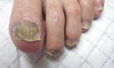
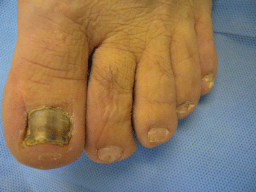
Fig. 3.2
DLSO in a diabetic patient. All nails are affected. Note the tinea pedis scaling and the hematoma of the great toe (due to poor fitting shoes, which is common due to neuropathy)
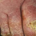
It’s particularly important to keep in mind the relationship between DLSO and tinea pedis [7]. The same dermatophyte organism (Trichophyton rubrum) is the major cause of both fungal infections [10, 27], and therefore each condition can serve as a reservoir of infection for the other. That’s why DLSO is almost always preceded (or accompanied) by an infection of tinea pedis (Fig. 3.3). This relationship partially explains the greatly increased frequency of toenail compared to fingernail DLSO: the greater incidence of tinea pedis over tinea manuum provides more opportunities for the toenails to be infected.
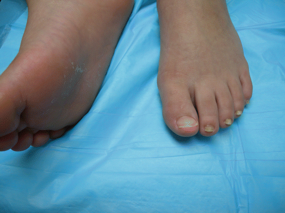
Fig. 3.3
DLSO and concomitant tinea pedis on the plantar surface of the opposite foot due to T. rubrum
Of course, if patients scratch or pick at their fungally infected feet, toes, or toenails, it’s possible for them to transfer the fungus onto their hands, which can cause them to develop tinea manuum and/or fingernail DLSO (Fig. 3.4). This often causes the patient to develop a relatively common pattern of infection known as “two feet-one hand syndrome (Fig. 3.5)” [28, 29].
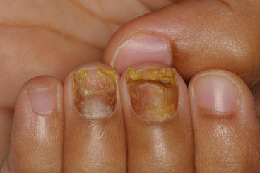
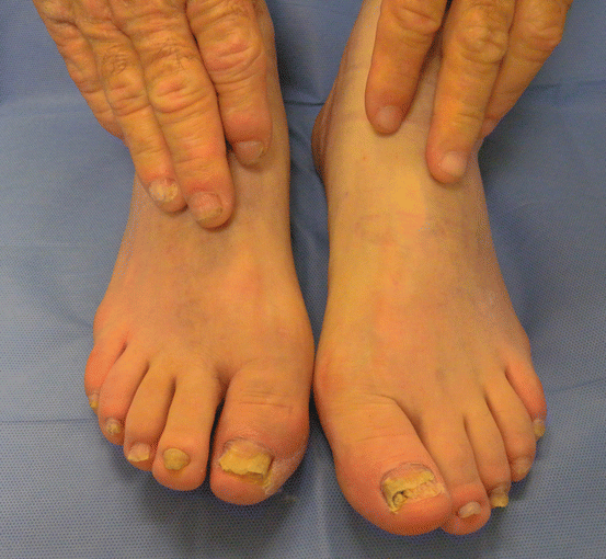
Fig. 3.4
An example of fingernail DLSO
Fig. 3.5
DLSO is present on the nails of both feet and the right hand, while the left hand is unaffected (two feet-one hand syndrome)
Overall, the incidence of DLSO is projected to continue increasing, largely due to a surge in critical risk factors like the age of the population, along with an increased prevalence of diabetes and peripheral vascular disease.
Clinical Features
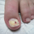
The characteristic features of DLSO are discoloration of the nail, subungual hyperkeratosis, and onycholysis (Fig. 3.6).
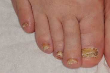
Fig. 3.6
DSLO. Note the yellow discoloration, severe subungual hyperkeratosis, and onycholysis
True to its name, DLSO’s fungal organism invades the nail bed at the distal portion of the nail. The fungus usually first infects the palm or sole, causing tinea pedis or tinea manuum, and then spreads from the skin to the nail bed via the hyponychium or the lateral nail fold.
In the early stages of infection, the fungus is limited to the nail bed, and the nail plate may appear normal. During this time, the stratum corneum of the nail bed often begins to thicken (subungual hyperkeratosis) due to mild inflammation from the fungal infection. The resulting keratotic debris pushes the nail plate up, eventually causing onycholysis.
From the initial site of infection, fungi migrate proximally (toward the cuticle) along the longitudinally oriented rete ridges of the nail bed. This explains the yellow, orange, or white longitudinal spikes and streaks which are a typical sign of progressing disease (Figs. 3.7 and 3.8).
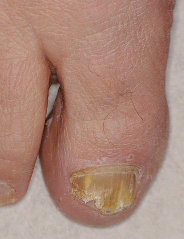
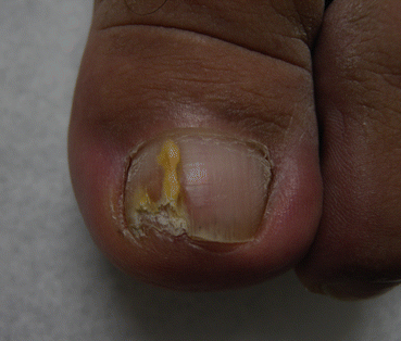
Fig. 3.7
The proximal progression of the fungus causes the yellow streaks leading toward the proximal nail fold. Also, note the tinea pedis scaling
Fig. 3.8
Another example of a discolored longitudinal streak due to proximal fungal progression, this one extending all the way into the lunula. There is also subungual hyperkeratosis
The nail plate may also display diffuse discoloration and look yellow, orange, or white. Less commonly, when DLSO is caused by non-dermatophytic fungal molds that produce melanin, the nail presents with a brown/black pigmentation (ungual melanonychia) similar in appearance to nail melanoma (Fig. 3.9). Thankfully, this is relatively rare, as many of the organisms which cause ungual melanonychia (e.g., Neoscytalidium dimidiatum) do not respond to antifungal therapies and are very difficult to cure [30].