© Springer International Publishing Switzerland 2017
Antonella Tosti, Tracey C. Vlahovic and Roberto Arenas (eds.)Onychomycosis10.1007/978-3-319-44853-4_2020. Onychomycosis: Procedures and Laser Treatment
(1)
Impression Foot & Ankle, 201 W. Guadalupe Rd. Ste. 318, Gilbert, AZ 85233, USA
(2)
Department of Podiatric Medicine, Temple University School of Podiatric Medicine, 148 N 8th Street, Philadelphia, 19107, PA, USA
Introduction
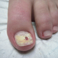
Onychomycosis is a very common problem in the podiatric practice with a reported incidence of 3.22 % in the general population, ranging from 0.14 % in children to 11.93 % in at-risk populations such as dialysis patients [1]. Current therapies have mostly focused on oral antifungals and, more recently, topicals such as efinaconazole and tavaborole. It is true that the oral antifungals have repeatedly proven to be the most effective for toenail fungus, and the newer topical medications’ increased efficacy is certainly encouraging. However, questions of their usefulness arise in patients in which these modalities are not affordable and/or plausible with concomitant medical issues. In these patients the question is: are there other modalities for treating nail fungal infections, whether it be distal lateral subungual onychomycosis or total dystrophic onychomycosis?
Recently that answer is in the affirmative with new modalities coming to market in the last few years. In the past, that answer involved surgical intervention with or without concomitant antifungal treatment. Various surgical methods were studied, including debridement, abrasion, avulsion, matrixectomy, and more. More recently, laser and light therapy has become an area of interest for researchers. This chapter will focus on these alternative therapies and discuss the latest research on their application and efficacy.
Procedures
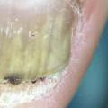
Just as in bacterial infections in the diabetic foot, frequently surgical correction of a fungal infection is a viable option. Many already do this as a part of “routine nail care,” which in the case of fungal nail infections can be considered a surgical removal. We may think we’re simply trimming nails for patients’ comfort, but we are also decreasing fungal burden as we remove those thick brittle distal nail portions, just as if we were removing the overlying bacterial contaminants in a wound debridement. In cases of very limited onychomycosis, such as a dermatophytoma within the nail or early distal lateral subungual onychomycosis, this may be enough to remove the entire fungal burden.
However, most of the time simple nail debridement is not enough to achieve complete fungal cure. Concomitant oral or topical antifungal therapy is usually required to completely eradicate the infection. Malay et al. in 2009 studied 55 patients with onychomycosis and compared “routine nail care” alone to nail debridement combined with topical antifungal therapy. Not surprisingly, none of the patients in the debridement-alone group achieved mycological cure while 76.74 % of those receiving combined therapy did [2]. Ameen et al. in 2014 also recommended adding antifungal therapy to debridement but weren’t optimistic about its results even with avulsion due to a disappointing randomized controlled trial that came out about the same time [3].
Some have suggested that abrasion or nail drilling, rather than debridement, may be just as successful in eradication of the fungal infection. Tchernev et al. suggested surgical removal for resistant infections but recommended abrasion therapy or superficial scraping of the nail plate if possible [4]. On the contrary, Cursi et al. studied the efficacy of oral and topical antifungals combined with nail abrasion in 2013 [5]. They achieved only 32–36 % complete or partial cure in 25 patients after 12 months with 24 % recurrence even in those who did achieve cure. More studies need to be done in this regard.
Sometimes debridement even with antifungal therapy is not enough, though, and more drastic surgical procedures need to be done, such as avulsions or matrixectomies. Regarding avulsions, painful pressure due to a thick mycotic nail can easily be relieved with temporary removal of all or part of the nail, but concomitant topical or oral antifungal therapy is also required here or the result will be as short lived as the procedure [6]. Moreover, total nail avulsions should be avoided as they can lead to shrinkage of the nail bed, distal in-growing edge, and upward growth of the nail bed due to the loss of compressing forces of the nail plate [7]. Baran et al. in 1985 achieved total cure in 20 patients treated with partial nail avulsions combined with topical and systemic antifungal therapy; however, it took 6–18 months [8]. Lai et al. performed 33 nail avulsions in 32 patients more recently and achieved complete cure or almost complete cure in 88 % of patients, but these patients also received topical or systemic antifungal therapy [9].
When avulsions combined with antifungal therapy fail to provide relief, permanent matrixectomy should be considered. Despite popular belief, there are multiple ways of doing this. The most popular technique involves removal of all or part of a nail with the matrix attached and afterward applying a chemical such as 89 % phenol or 10 % sodium hydroxide to the matrix to prevent regrowth. Proponents of sodium hydroxide say that it has a higher success rate with less drainage and faster healing times [6]. In either case, a tourniquet should be used as any blood in the surgical field will dilute the chemical and decrease effectiveness. The chemical of choice is usually applied three to four times for 30 s each, after which it is rinsed with alcohol (in the case of sodium hydroxide, it is neutralized due to an acid-base reaction). Care should be taken to apply the chemical not only to the matrix but to the nail bed in order to get the ventral nail matrix and prevent regrowth.
Other methods of removing the nail exist but are much less frequently used. These include negative galvanic current, radio-wave electrical energy, carbon dioxide lasers, occlusive urea, and “cold-steel” procedures such as the Winograd, Frost, and Zadik nail procedures. Negative galvanic current produces heat and production of sodium hydroxide through electrolysis at the cathode that is applied to the matrix for 4–7 min. Frequent users of this technique report decreased post-op pain, inflammation, and drainage than chemical matrixectomy [6]. Radio-wave electrical energy was described by Hettinger et al. in 1991 and is applied directly to the matrix via a probe. It only takes 2–4 s and provides matrix destruction and hemostasis by an electrocautery effect. Users report less postoperative pain, swelling, and infection than other methods, but no data supports this. Obviously this method cannot be used in people with pacemakers [6]. So-called “cold-steel” procedures such as the Frost and Zadik are good for truly resistant cases, and they give the added benefit of access to exostoses needing removal.
Related posts:

Stay updated, free articles. Join our Telegram channel

Full access? Get Clinical Tree


