Chapter 75 Numbness/Saphenous Nerve
Introduction
Leg numbness due to nerve damage is one of the considerable complications after anterior cruciate ligament (ACL) reconstruction using both bone–patellar tendon–bone (BPTB) and medial hamstring tendons. Such patients especially complain of uncomfortable feelings when falling on their knees. Harvesting a BPTB graft includes risks of damaging nerves and causes sensory disturbance.1,2 Pagnani et al3 pointed out the risk of saphenous nerve damage by harvesting medial hamstring tendons in the region of the pes anserinus.
Many authors have reported the nerve distribution patterns of the infrapatellar regions.2,4–12 It is well known that both the medial cutaneous nerve of the femoral nerve and the infrapatellar branch of the saphenous nerve are distributed throughout the infrapatellar region and the anterior lower leg region.9,10,12 The saphenous nerve descends laterally along the femoral artery and enters the adductor canal. It then leaves the artery at the distal end of the canal to proceed vertically along the medial side of the knee and runs between the sartorius and gracilis tendons. In contrast, the medial femoral cutaneous nerve originates from the anterior cutaneous branches of the femoral nerve. The medial femoral cutaneous nerve runs laterally to the femoral artery, and then it crosses anteriorly to the artery at the apex of the femoral triangle to be distributed to the anteromedial thigh and the infrapatellar region. Branches of the medial femoral cutaneous nerve and the infrapatellar branch of the saphenous nerve connect to each other9,10,12 and form the subsartorial plexus in the infrapatellar region.13
Bone–Tendon–Bone Autograft
Anterior knee pain including leg numbness has been reported as a main complication of ACL reconstruction using BPTB grafts. In previous reports, the rate of postoperative anterior knee pain ranged from 4% to more than 40%.8,14,15 Mishra et al at first described a technique using two horizontal incisions for patellar tendon harvest for the purpose of more cosmetic scarring and reducing pain and flexion limitation.16 Kartus et al17 changed to two vertical incisions and reported of an insensitive area compared with the insensitive area that resulted from a traditional vertical incision, which averaged 24 cm. Tsuda et al18 changed the method of approaching the retinaculum layer, opening it horizontally rather than splitting it to protect nerves using two horizontal incisions, and they reported a 17% rate of postoperative leg numbness. Portland et al19 compared a horizontal incision and a vertical incision and reported a infrapatellar numbness of 43% resulting from a horizontal incision and 59% resulting from a horizontal incision.
Hamstring Autograft
The donor site morbidity associated with harvesting a hamstring tendon graft is well recognized to be less common than that associated with harvesting a BPTB autograft.20 However, sensory disturbance is frequently observed in regions on the anterior lower leg after ACL reconstruction using medial hamstring tendons.21,22
Clinical Examination
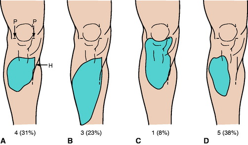
We clinically examined 103 patients who had arthroscopically assisted ACL reconstructions using medial hamstring tendons to investigate the frequencies and areas of sensory disturbance.23 As an operative procedure, we made two horizontal incisions for the arthroscopy portal and one longitudinal incision (2.5–3 cm) at the pes anserinus for the tendon harvest and the tibial drill holes. We performed an inside-out technique and used Endobutton (Smith & Nephew Endoscopy, Andover, MA) for femoral fixation. The clinical examination was performed for an average of 13 months (range 6–18 months) after the operation. We detected sensory disturbance on the anterior surface of the lower leg in 60 of 103 (58%) patients. We randomly selected 13 patients with sensory disturbance and neurologically examined in detail the regions of sensory disturbance. The regions of sensory disturbance were of various sizes and shapes (Fig. 75-1). These regions were generally quadrilateral and located lateral to the longitudinal incision for tendon harvest and distal to the horizontal incisions for the arthroscopy portal. In the detailed neurological examination, in 8 of 13 legs (62%) the region was very close to the longitudinal incision (see Fig. 75-1, A to C), and in the other 5 legs (38%) it was relatively far away from the longitudinal incision (see Fig. 75-1, D). The region was lower than the superior end of the longitudinal incisions in 12 of 13 legs (92%); however, in one leg (8%), the region was close to the horizontal incisions (see Fig. 75-1, C). The region was located in the upper half of the lower leg in 10 legs (77%); however, in three legs (23%), the region was wider than others (see Fig. 75-1, B).
Anatomical Investigations
In our anatomical study, 51 lower limbs of 26 adult cadavers were used.24 In the mediodistal region of the patella, the nerve branches pierced the fascia cruris in various patterns and ran on the outer surface of the fascia to supply the skin (Fig. 75-2). On the outer surface of the fascia, the nerve branches often were connected to each other. In the regions near the horizontal and longitudinal skin incision lines, the nerve branches ran on the outer surface of the fascia cruris in all legs.
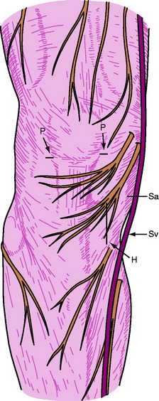
Fig. 75-2
Related posts:
 Anatomical Anterior Cruciate Ligament Reconstruction with Double-Bundle, Double-Stranded Hamstring Autografts: A Four-Tunnel Technique
 Tibial Fixation for Anterior Cruciate Ligament Hamstring Grafts: 10 Techniques that Improve Fixation
 Hamstring Regeneration Following Harvest for Anterior Cruciate Ligament Reconstruction: A Review of the Current Literature
 Anatomical Double-Bundle Reconstruction of the Anterior Cruciate Ligament
 Stiffness: Prevention and Treatment
 Endobutton Anterior Cruciate Ligament Reconstruction Femoral Fixation
Stay updated, free articles. Join our Telegram channel

Full access? Get Clinical Tree


