Chapter 25 Anatomical Double-Bundle Reconstruction of the Anterior Cruciate Ligament
Introduction
Anterior cruciate ligament (ACL) reconstruction remains one of the most common procedures performed by orthopaedic surgeons in the United States, with approximately 100,000 performed per year.1 ACL surgery has evolved tremendously from the original open techniques to modern procedures focusing on endoscopic reconstruction of the anteromedial (AM) bundle using a variety of graft choices and fixation techniques. However, the success of single-bundle ACL reconstruction ranges from 69% to 90%.2,3 In addition, according to Fithian et al,4 95% of patients who underwent single-bundle ACL reconstruction developed medial compartment degenerative radiographic changes after 7 years, and only 47% were able to return to their previous activity level. Because arthrosis was observed medially, it could not be attributed to the initial subluxation event, which usually results in a bone contusion or a concomitant meniscal tear involving the lateral compartment.4
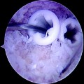
Single-bundle ACL reconstruction is the “gold standard,” but some authors have noted persistent instability with functional testing of single-bundle ACL reconstruction.5,6 Thus, there is a growing trend toward a more anatomical ACL reconstruction that recreates both the AM and the posterolateral (PL) bundles. The double-bundle anatomy of the ACL was first described in 1938 by Palmer et al.7 The terminology of the AM and PL bundles are chosen according to their tibial insertions. The tibial and femoral insertion sites of both the AM and PL bundles have been well described.8,9 The femoral origin has an oval shape, with the center of the AM bundle close to the over-the-top position and the center of the PL bundle close to the anterior and inferior cartilage margin. The femoral origin site changes as the knee is taken through an arc of motion. The two bundles are parallel with a vertical orientation when the knee in extension (i.e., the AM footprint is situated directly superior to the PL footprint). This changes to a more horizontal orientation, with the PL footprint becoming actually anterior to the AM footprint when the knee is flexed beyond 90 degrees. The changing orientation of the two bundles’ footprints as the knee is taken through an arc of motion leads to the observed crossing pattern of the independent components of the ACL. Although the two bundles are intertwined, their functional tensioning pattern is independent throughout the knee’s range of motion.10 Close to extension, the AM is moderately loose and the PL is tight. As the knee is flexed, the femoral attachment of the ACL takes a more horizontal orientation, causing the AM bundle to tighten and the PM bundle to loosen. The ACL has been described as a restraint to anterior tibial displacement and internal tibial rotation. The rotational stabilizing component might be better attributed to the PL bundle.
The idea of reconstructing both bundles of the ACL was described by Mott and Zaricznyj in the 1980s.11,12 They independently described a double-bundle technique. Mott drilled two separate tunnels, whereas Zaricznyj used a single femoral and two tibial tunnels. Despite publishing their results, the technique did not become mainstream. Recent biomechanical evidence supports the anatomical double-bundle ACL reconstruction as more accurately recreating the natural anatomy.13,14 Both translational and coupled rotational translation were significantly less in the specimens with double-bundle ACL reconstruction. We present the senior author’s (F.H.F) technique of anatomical double-bundle ACL reconstruction with two femoral and tibial tunnels using two tibialis anterior allografts.
Preoperative Considerations
History: Signs and Symptoms
ACL injuries occur frequently in sports that involve running, jumping, and cutting movements. They can occur without contact when the foot is anchored to the playing surface—usually by way of cleats or a rubber sole—and the body rotates beyond the tolerance of the ligament as the knee buckles. Thus, it is important to ask the patient how the injury occurred and the position of the knee during the injury, which may also allude to the ACL bundle injury pattern.15 This may be associated with an audible “pop.” Asking whether the athlete was able to continue to play will give you an idea of the severity of the injury. Knee pain and a hemarthrosis are usually present acutely. A complaint of instability is also common, especially with walking downhill or down stairs.
Indications
The absolute indications for double-bundle ACL reconstruction are evolving. Even though single-bundle ACL reconstruction is considered the “gold standard,” the technique can be improved. Gait analysis after single-bundle reconstruction has demonstrated that rotatory instability persists.5 Furthermore, biomechanical cadaveric studies have shown that even lowering the femoral insertion site to the 3- or 9-o’clock position does not fully prevent rotatory instability.16 Clinically, as many as one-fifth of the patients do not resume preinjury activities and usually complain of vague instability symptoms that objectively correspond to a mild persistent pivot shift.17 In comparison, double-bundle ACL reconstruction does restore the rotational component in a cadaveric model.14 It has been suggested that a positive pivot shift after ACL reconstruction is correlated with the development of later osteoarthrosis.18 Perhaps with reconstruction of both the AM and PL bundles, the decreased rotational instability will provide improved overall knee kinematics and may prevent or slow the degenerative changes seen after single-bundle ACL reconstruction.4 A contraindication to performing the double-bundle technique is in the young athlete with open physes. Two tunnels would risk physeal arrest with subsequent malalignment and possible leg length discrepancy.
Surgical Technique
Anesthesia and Positioning
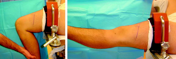
The operative extremity is identified by the patient and initialed by a member of the surgical team. All patients undergo a preoperative femoral nerve block in the holding area by our anesthesia colleagues. The patient is then placed in a supine position and given intravenous conscious sedation. A careful exam under anesthesia is performed and recorded to document the Lachman and pivot-shift maneuvers. Again, the senior author is interested in correlating the exam with the tear pattern of the individual bundles of the ACL. A tourniquet is applied to the proximal thigh. The extremity is then secured within a circumferential leg holder placed at the level of the tourniquet. The foot of the operating table is completely retracted to permit hyperflexion of the knee, which is crucial for later placement of the PL femoral tunnel. The contralateral extremity is placed within a well-leg holder with the hip flexed approximately 90 degrees and abducted and externally rotated away from the surgical field to allow unobstructed access to the operative knee (Fig. 25-1). The leg is elevated for 5 minutes, and the tourniquet is then inflated. The knee is prepped and sterilely draped.
Anterior Cruciate Ligament Graft Preparation
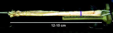
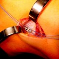
Two tibialis anterior allografts are individually fashioned as a double loop. The folded length of each graft should be approximately 12 cm for sufficient graft tissue. The grafts are trimmed to a folded diameter of 7 mm for the PL bundle and 8 mm for the AM bundle. A #2 braided suture is whipstitched up and down both ends of the graft for 3 cm. The stitch depth is alternated, and care is taken to avoid penetrating the suture and risking weakening or breaking. The graft is then passed through the closed-looped Endobutton (Smith & Nephew, Andover, MA). Two Fiberwire sutures (Arthrex, Naples, FL) (one stripped and one nonstripped for later identification) are placed within the button holes. A 2–0 absorbable suture is tied through both strands of the folded graft to secure them once the graft is passed within the closed-looped Endobutton. Each graft is marked to alert the surgeon when to engage or “flip” the Endobutton (Fig. 25-2).
Surgical Landmarks
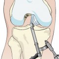
With the knee flexed approximately 45 degrees, the inferior pole of the patella is marked. The inferior extent of the lateral parapatellar portal begins at the level of the inferior pole of the patella and extends proximally for approximately 2 cm. The medial parapatellar portal begins at the level of the inferior pole of the patella and extends distally along the medial aspect of the patellar tendon for approximately 2 cm. The high placement of the portals allows the arthroscopic instruments to enter the knee above the level of the fat pad. The 11 scalpel blade is angled approximately 45 degrees to the skin and distally toward the notch to safely enter the knee without harming the articular cartilage. A low AM accessory portal will be placed with the assistance of a spinal needle to ensure proper trajectory for the PL femoral tunnel (Fig. 25-3
Related posts:
 Anatomical Anterior Cruciate Ligament Reconstruction with Double-Bundle, Double-Stranded Hamstring Autografts: A Four-Tunnel Technique
 Hamstring Regeneration Following Harvest for Anterior Cruciate Ligament Reconstruction: A Review of the Current Literature
 Revision Anterior Cruciate Ligament Reconstruction Using Autologous Hamstring Tendons
 High-Stiffness, Slippage-Resistant Cortical Fixation Has Many Advantages over Intratunnel Fixation
 The Retrodrill Technique for Anterior Cruciate Ligament Reconstruction
 Endobutton Anterior Cruciate Ligament Reconstruction Femoral Fixation
Stay updated, free articles. Join our Telegram channel

Full access? Get Clinical Tree


