Major surgical body contouring procedures have several inherent drawbacks, including hospitalization, anesthetic use, pain, swelling, and prolonged recovery. It is for these reasons that body contouring through noninvasive and minimally invasive methods has become one of the most alluring areas in aesthetic surgery. Patient expectations and demands have driven the field toward safer, less-invasive procedures with less discomfort, fewer complications, and a shorter recovery. In this article, the current minimally invasive and noninvasive modalities for body contouring are reviewed.
Key points
- •
The strong patient demand for safer, less invasive body contouring procedures will inevitably drive technology toward more effective modalities that minimize downtime and recovery, while improving results.
- •
The future of body contouring will likely stratify patients into more distinct categories, such as those requiring aggressive surgical excision, minimally invasive liposuction techniques with adjunctive energy-delivering modalities or mesotherapy, and noninvasive techniques achieving volume reduction and skin tightening through additional energy-delivering modalities.
- •
Currently, the most ideal candidates for these types of procedures are those who are accepting of a mild to moderate result.
Minimally invasive modalities
Introduction
Surgical body contouring procedures have several inherent drawbacks, including hospitalization, anesthetic use, pain, swelling, and prolonged recovery. It is for these reasons that body contouring through noninvasive means has become one of the most alluring areas in aesthetic surgery. Patient expectations and demands have driven the field toward safer, less-invasive procedures with less discomfort, fewer side effects, and a shorter recovery.
The future of body contouring will most likely involve completely noninvasive procedures for mild cases, minimally invasive procedures for moderate cases, and invasive procedures reserved for massive weight loss and larger patients.
In this article the current minimally invasive and noninvasive modalities for body contouring are reviewed.
Adjunctive modalities in liposuction
After the modern reinvention of liposuction more than 30 years ago, liposuction was performed as an inpatient procedure, often requiring blood transfusion postoperatively. The introduction of the tumescent technique has significantly optimized the outcomes and safety profile of liposuction procedures and has subsequently become the gold standard in body contouring procedures. Furthermore, refinement of body site-specific cannulas and the use of manual syringe suction for autologous transfer and fine contouring have optimized liposuction techniques and improved outcomes. Adjunctive energy-delivering modalities, such as laser-assisted liposuction (LAL) and ultrasound-assisted liposuction (UAL), have shown promise in facilitating fat removal, reducing procedure duration, surgeon strain, patient recovery time, and postoperative pain.
Laser-Assisted Liposuction (LAL)
LAL uses a small fiber (300 to 1000 μm) delivered through a narrow cannula of approximately 1 mm to deliver energy to tissues. The distinct advance in laser delivery has allowed for the delivery of laser energy under, rather than through, the skin. This approach allows more energy to be placed directly at the target instead of passing through epidermis and dermis, with simultaneous cooling mechanisms for protection of the surface from intense heat. With the safety mechanism defined and delineated, the advantage of the laser is the proven efficacy in skin tightening. An additional advantage of LAL is the ability to treat scarring, dimpling, and cellulite in the superficial layers with the small cannula. This small cannula reduces the risk of contour irregularities seen with larger cannulas. Some disadvantages include the higher cost of equipment, the need for precise temperature measurement, and the potential for burns or blisters without this monitoring. Previous disadvantages were related to duration of the procedure; however, as technology progresses, additional wavelengths have been proven to be 40 times more efficient in fat disruption while maintaining similar cannula size.
Preoperative preparation
Patients are evaluated for potential fat reduction and skin laxity improvement. When treating specific areas, these areas of adiposity are marked in a standard fashion, in addition to 5 × 5 cm squares representing the areas of skin laxity targeted for energy application. Approximately 50 to 100 mL of tumescent fluid is administered per 5 × 5 cm sector ( Fig. 1 ). This procedure is routinely performed with oral and local anesthesia only, but may be supplemented with intravenous (IV) sedation, epidural block, or general anesthesia.
It is essential to manage patient expectations during the initial consultation. Patients must understand that the skin tightening effect hinges on several variables, including age, genetics, and skin condition from environmental factors, such as smoking and sun exposure.
Surgical technique
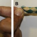
The laser system (Triplex; Cynosure, Westford, MA, USA) allows individual as well as sequential emission of 1064-nm, 1320-nm, and 1440-nm wavelengths. Energy is delivered to the subdermal tissue through a 600-μm or 1000-μm fiber threaded through a 1-mm microcannula that extends 2 to 3 mm beyond the distal end of the microcannula. When the microcannula is inserted into the tissue, the laser is activated and the microcannula is moved slowly and evenly through the deep fat layer with a 1064-nm to 1440-nm combination, or the superficial subdermal layer with a 1064-nm to 1320-nm combination ( Fig. 2 ). For ideal absorption and performance, wavelength combinations are used for preferential target affinity of fat and water, respectively.
Using the biplanar technique, adipose cells are disrupted, resulting in a more liquefied material that can be removed through a smaller cannula. This smaller cannula refines the removal, particularly close to the surface, thereby reducing potential contour irregularity that can be seen with larger instrumentation. To achieve increased skin elasticity and tightening, surface temperature goals of 40°C to 42°C superficially, and deep temperature goals of 45°C to 47°C are delineated. The lower temperature goal is used for darker skin colors. Achieving and maintaining these temperature goals uniformly is essential to achieving optimal efficacy.
Safe energy delivery is monitored via an accelerometer delivery system (Smart Sense; Cynosure) attached to the laser handpiece, used to minimize the occurrence of localized thermal damage during treatment. The handpiece contains a motion-sensing feedback chip that provides constant information of cannula movement to the laser device. Slower movement of the handpiece triggers a reduction in laser power. If the handpiece stops, energy delivery ceases within 0.2 seconds. This safety feature prevents excess accumulation of energy in a given area. Temperature safety is measured with a thermastore probe at the tip of the cannula, which is set for a safe range according to the above-mentioned parameters. When the threshold is reached, the laser automatically stops firing.
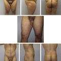
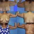
A 2-layer/2-step technique is used in which the deep fat layers (1–3 cm below the epidermis) within the premarked squares are treated first (see Fig. 2 ). Insertion of the microcannula is facilitated with the use of 1-mm incisions made with a number 11 blade for appropriate access to the treatment area. The superficial subdermal layer (0.5 cm below the epidermis) is treated in the second step of the 2-layer technique ( Fig. 3 ). Aspiration is performed with a standard 3-mm suction cannula to remove any remaining fat, disrupted cells, and free fat oils. Clinical examples are shown in Figs. 4–6 .
Optimization of this procedure hinges on proper delivery of energy while maintaining an appropriate safety margin. Inadequate tightening can often be the result of insufficient temperature attainment necessary to achieve the desired effect. Too much energy applied in one area can result in excessive heat build-up and fat necrosis. If too much heat is applied superficially, the result will be skin blistering, burns, or pigmentation changes. If used properly, the built-in safety mechanisms will help control temperature and avoid harmful buildup of heat.
Postoperative care
Firm pressure dressings are applied to the wounds on completion of the aspiration, and compression garments are worn for 3 to 4 weeks postoperatively. Dependent incisions are left open for drainage, and oral antibiotic prophylaxis is administered.
Skin and tissue firmness is examined 1 week postoperatively. Excessive swelling or dense edema is treated with the use of the Triactive Device (Cynosure) or equivalent massage, edema reduction, and lymphatic drainage enhancement for 3 weeks postoperatively. Patients are reminded of the time necessary for skin changes to occur during the dynamic fibroblast stimulation period (90 days). If further enhancement of skin is desired after 3 months, acceptable external skin devices can be applied, and additional treatments in adjacent areas can be performed after tissue effects have subsided.
Complications and management
Complications of LAL include blisters, end hits, burns, blowholes, prolonged edema, heat/pressure problems, neurapraxia, permanent nerve damage, contour irregularity, minor asymmetries, and insufficient effect. If performed properly, the occurrence of these complications is rare.
Summary
Previous work has led to an improvement in the safety and an enhancement of the cosmetic results of LAL. By reducing blood loss, minimizing complications, and promoting skin tightening, LAL has proven to be a safe and effective body contouring procedure in appropriate candidates. Future applications of fiber-deliverable energy will aim to address the superficial irregularities of the skin, such as dimpling, scars, and cellulite.
Ultrasound-Assisted Liposuction (UAL)
Ultrasonic energy was first applied to body contouring surgery in the late 1980s by Scuderi and coworkers and later popularized by Zocchi, who introduced the technique to the United States in 1993. The third-generation VASER system (Sound Surgical Technologies, Louisville, CO, USA) was introduced in 2001 and is currently the most commonly used UAL technology.
Ultrasound interacts with tissue by 3 basic mechanisms: thermal, mechanical, and cavitation. The thermal effect is initiated by the heat generated from the rapidly vibrating ultrasonic probe. The mechanical effect is created when the rapidly vibrating tip of the ultrasonic probe contacts tissue. Cavitation is the tissue interaction most responsible for causing fat emulsification with current UAL devices. When the wetting solution is dispersed within the tissues, small microbubbles are lodged within the fat tissue matrix, which then implode and collapse with the application of ultrasound. This process separates fat cells within the fat tissue matrix, which subsequently mix with the tumescent solution by means of acoustic streaming to create an emulsion. This emulsion is subsequently harvested by means of a suction cannula.
Preoperative preparation
Even though UAL has expanded the parameters for liposuction patient selection, one criterion that remains unchanged is the patient’s candidacy to undergo an elective surgical procedure. Once this has been established, preoperative preparation may proceed.
Preoperative markings are performed in the standing position. There are 5 distinct anatomic areas that should be avoided in patients undergoing UAL. These areas, termed the “zones of adherence,” include the gluteal crease, the lower lateral thigh area of the iliotibial tract, posterior distal thigh above the popliteal crease, midmedial thigh area, and lateral gluteal depression area. Violation of these areas often leads to iatrogenic contour deformities. During the preoperative marking process, it is essential to plan the placement of access incisions carefully. UAL requires a greater number of slightly longer access incisions to accommodate the skin protectors and to avoid placing torque on the ultrasonic probe over curved anatomic areas.
VASER probe selection must consider the characteristics of localized fat in the region to be treated. The characteristics of fat cells in different regions of the body differ with respect to collagen structure and septi among the fat cells, as described by de Souza Pinto and colleagues. Knowledge of these differences is essential for probe selection to achieve optimal outcomes.
Penetration of tissue is influenced by probe diameter and the number of grooves at the tip. For a probe of a given diameter, more grooves emulsify fat tissue more efficiently. However, they do not penetrate fibrous tissues easily because of vibratory energy that is transferred to the sides of the probe as opposed to the front surface. Fibrous tissues are better addressed with probes with fewer grooves. Smaller-diameter probes also penetrate fibrous tissue more easily. The 3.7-mm probes achieve rapid debulking and contouring of medium to large volumes of soft to fibrous tissues. The number of grooves at the tip of the probe will vary according to the fibrous nature of the anatomic area. For treating smaller, soft to extremely fibrous localized fat deposits in sensitive areas, fine contouring is achieved with 2.9-mm probes.
In general, continuous mode should be used for fibrous tissue, faster fragmentation, and times when tissue emulsification is not readily achieved in VASER mode. VASER mode is more suited for delicate work, finer sculpting, or softer tissues. The device must be adjusted such that the probe moves smoothly through tissue.
Experience and practice have delineated application times. In general, 1 minute of application time may be used per 100 mL of infused solution in VASER or continuous mode. Loss of resistance to probe movement in all intended areas can be considered the surgical endpoint. Following emulsification, aspiration can be performed with suction-assisted or power-assisted liposuction.
Surgical technique
The prone position provides good access to the back, flanks, lateral thighs, and superior posterior thighs. General endotracheal anesthesia is preferred for patients requiring prone positioning and for large volume aspirations. Many surgeons also prefer the lateral decubitus position, which requires one additional patient repositioning. Despite additional repositioning, some authors maintain that the lateral decubitus position offers better access with less trauma and is particularly helpful for evacuating large volumes from the flanks and back with the goal of creating more aesthetic waistlines. Supine positioning offers access to the abdomen, anterior and medial thighs, knees, calves, arms ankles, breasts, and face.
Maintaining core body temperature is best accomplished by running IV fluids through a fluid warmer in addition to the use of forced warm air by means of a Bair Hugger (Arizent Inc, Eden Paris, MN, USA). The access incisions are placed in previously marked areas using a number 11 blade and must be long enough to accommodate the fluid-infiltrating cannula. The rate of flow is controlled on the infusion pump according to the anatomic area. Generally, a wetting solution is prepared with 1 mL of epinephrine added to 1 L Ringer’s lactate at room temperature (1:1,000,000 dilution). In cases not using general anesthesia, lidocaine may be added to the wetting solution. The dose of lidocaine should not exceed 35 mg/kg, although some authors routinely use doses exceeding 50 mg/kg while maintaining a safety margin.
Treatment recommendations
Posterior trunk
- •
3.7-mm 2-ring probe at 80% energy level in continuous mode for most of the back
- •
3.7-mm one-ring probe at 80% energy level in continuous mode for tight fibrous back rolls
- •
Posterior trunk areas require slightly longer ultrasound application, usually between 12 and 14 minutes on average
- •
Aspiration is accomplished with a small-diameter cannula
Abdomen
- •
3.7-mm 3-ring ultrasonic probe is used at an energy setting of 80% in VASER mode
- •
Continuous mode is used for the area above the costal margin
- •
Average ultrasound time is 8 to 9 minutes
- •
Aspiration is accomplished with a small-diameter cannula
Extremities and buttocks
- •
3.7-mm 3-ring ultrasonic probe is used at an energy setting of 70% in VASER mode for the superior medial thigh
- •
Around the knees and superior posterior thigh, a short 3-mm 3-ring probe is used in 80% VASER mode
- •
Depending on how fibrous the subcutaneous layer may be, the anterior and lateral thigh areas are also performed with the 3.7-mm 3-ring probe in 80% VASER or continuous mode
- •
Average ultrasound time is approximately 3 minutes for both knees, 5 minutes for both superior medial thighs, 8 minutes for both anterior thighs, 6 to 7 minutes for both lateral thighs, and 4 minutes for both superior posterior thighs
- •
For the axillary area, a 3-mm, 3-ring probe is inserted into the subcutaneous space through a small access incision in the axillary fold
- •
The energy setting is 70% VASER mode, applied for approximately 2 minutes per arm
- •
Aspiration is performed through a small-diameter cannula
Gynecomastia
- •
3.7-mm one-ring probe or the gynecomastia arrow probe can be used efficiently in this area
- •
Energy levels are set at 80% to 90% at continuous mode at an average of 3 to 4 minutes per breast
- •
Aspiration is performed through a small-diameter cannula
Face and neck
- •
Three access incisions are recommended: one behind each earlobe and one in the submental crease
- •
Smaller-diameter, highly precise instrumentation is used for facial UAL
- •
Ultrasound is applied by a 2.4-mm 3-ring ultrasonic probe at 50% to 60% energy levels in VASER mode for approximately 2 to 3 minutes
- •
Aspiration is performed with a fine cannula
HIV-associated cervicodorsal lipodystrophy
- •
3.7-mm one- or 2-ring probe is used depending on the fibrous nature of the area
- •
The energy setting is 80% continuous mode and the time will vary according to the volume of fat being treated
- •
Aspiration is performed with a fine cannula
Optimizing outcomes
- •
Apply the least amount of ultrasound energy necessary to obtain fat emulsification
- •
The clinical endpoint of ultrasound application is loss of tissue resistance against the probe
- •
Incision placement must be planned appropriately to facilitate access to the treatment area while avoiding torque on the ultrasonic probe
- •
Small-diameter cannulae provide the greatest precision, particularly in the aspiration of superficial fat
- •
Liberal use of wetting solution dispersed evenly through the fat maximizes efficiency of ultrasonic cavitation, minimizes blood loss, and provides added protection against thermal effects of UAL
- •
Circumferential contouring provides a more harmonious result than local fat extraction
- •
Postoperative use of foam compression garments, lymphatic drainage massage, and skin-moisturizing regimens can optimize UAL outcomes and decrease recovery times
Postoperative care
Following large-volume UAL procedures, close monitoring of fluid replacement and urine output is required. Postoperatively, most patients with large volume aspirations continue to leak fluid through the incisions for 24 to 36 hours. A significant volume of the infiltrating solution does get absorbed during the first 12 hours following major UAL and this must be considered when planning fluid replacement. Oral intake of fluids is permitted on waking. Early ambulation is encouraged, and patients are discharged on the first postoperative day. Foam and compression garments can be applied to most UAL patients in the immediate postoperative period. Lymphatic drainage and skin moisturizing regimens are helpful as soon as the patient can tolerate them.
Complications and management
The most common complications of liposuction procedures are under-extraction, over-extraction, or irregular contour. In general, these complications are prevented by the use of intraoperative flow sheets documenting infusion and aspiration volumes for each anatomic area. Under-extraction is usually corrected by revisionary extraction, whereas overcorrection may require fat grafting. Paresthesias, edema, and ecchymosis are usually self-limiting. The vibrating ultrasonic probe generates heat, which could lead to thermal injury, particularly around the incision site. The use of skin protectors is essential, along with the use of a wet towel adjacent to the incision to provide added protection. The most important factor in preventing skin burns is avoiding torque on the probes.
Seromas are the result of too much ultrasonic energy applied to the tissues either as a result of increased generator settings or prolonged application. It is seldom necessary to use energy settings greater than 80% applied for 1 to 1.5 min/100 mL of wetting solution to a particular area to achieve proper tissue fragmentation and emulsification.
Related posts:

Stay updated, free articles. Join our Telegram channel

Full access? Get Clinical Tree


