This article discusses management of the post–weight loss thigh deformity. Beginning with an explanation of the soft tissue variables contributing to the thigh and medial thigh deformity in the postbariatric individual, the article describes the important elements of selecting and screening candidates for surgery and the ideal sequence of procedures that should be followed to optimize results in this patient population. A detailed step-by-step description of the author’s technique for medial thigh lift is provided along with multiple examples of outcomes. Aftercare is reviewed along with potential complications and their management.
Key points
- •
Careful patient screening and education are critical to optimizing the outcome of medial lift procedures.
- •
Major variables contributing to the thigh deformity include descent of the soft tissues of the hips, lower abdomen and mons pubis; inferomedial migration of thigh soft tissues; descent of the buttocks; and circumferential thigh soft tissue excess secondary to weight changes and aging. Prior liposuction may also contribute to thigh contour deformities.
- •
An effective medial thigh lift requires prior correction of soft tissue excess along the lower body.
- •
Thigh lift procedures with an excisional component limited to the thigh perineal crease usually provide little if any additional benefit to individuals who have undergone an effective circumferential lower body lift procedure.
- •
Liposuction in vertical medial thigh lift procedures serves to create a readily identifiable and safe plane of dissection and to improve outcome by optimizing the amount of soft tissue that can be removed.
- •
A lower body lifting procedure and subsequent medial thigh lift with a vertical component help minimize the likelihood of the well-described complications associated with medial thigh lifts with an approach limited to the thigh perineal crease: labial spreading and scar migration.
Introduction
Thigh contour deformities, and in particular those of the medial thighs, are a frequent concern for individuals seeking body contouring. The deformity is usually secondary to weight loss and is often associated with prior pregnancy and, in many instances, liposuction of the thighs. Despite the frequency of this concern, plastic surgeons have often been reluctant to use the medial thigh lift procedure because of the risk for significant complications and poor results, and the potential for readily visible scars. Until recently, nearly all medial thigh lift techniques described in the literature attempted to address the medial thigh deformity by removing soft tissue excess in a vertical vector alone. An exception was the first description of a medial thigh lift in 1957 by Lewis. He advocated both a horizontal and vertical component to the procedure. Lockwood , a proponent of vertical vector excision alone, described minimizing complications such as labial spreading and scar migration and improving outcome by securing the medial thigh flap to Colles fascia. Although the concept seemed to be a logical approach to address problems associated with medial thigh lifts, it added little to existing procedures. In recent years, primarily in response to the increase in numbers of individuals with postbariatric body contour concerns, plastic surgeons have again advocated the excision of soft excess in both a vertical and horizontal vector to address medial thigh deformities. In addition, the significance of addressing the vertical soft tissue excess of the lower abdomen, hips, thighs, and buttocks before performing an effective medial thigh lift has become more generally accepted.
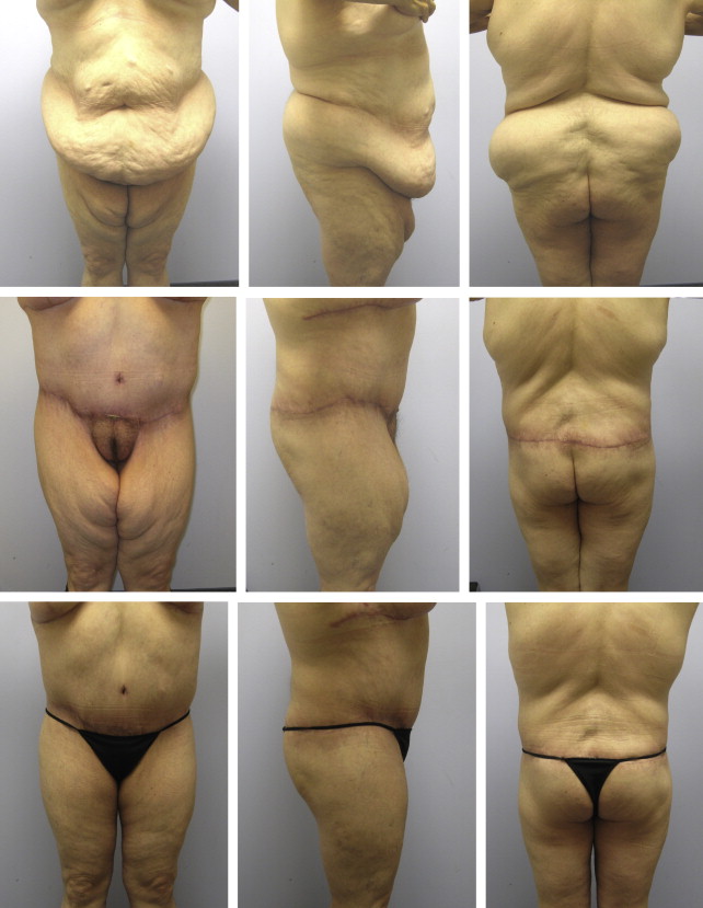
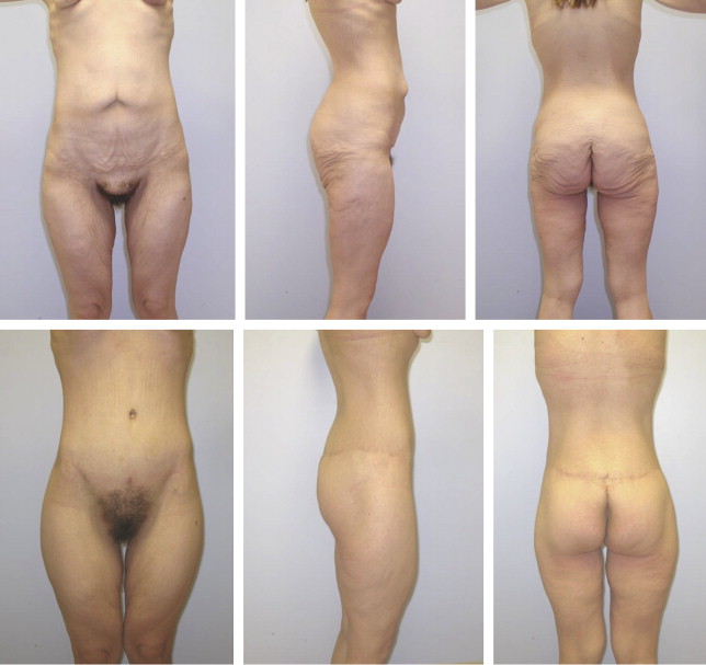
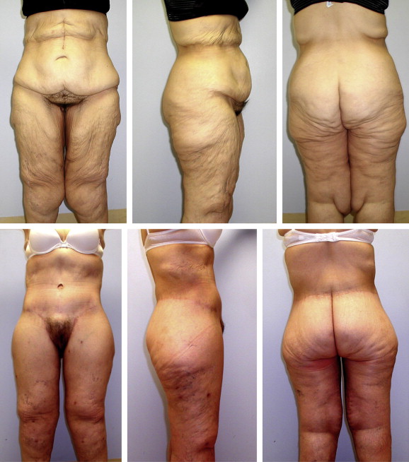
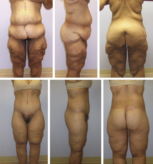
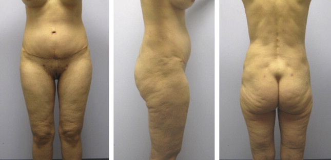
Our approach to medial thigh contouring is to address the variables outside the medial thighs affecting the medial thighs before performing a medial thigh lift procedure. In most instances, a body lift or simultaneous abdominoplasty, thigh lift, and buttock lift is performed first. For a small number of individuals, only an abdominoplasty is performed initially. For some patients, the need for a medial thigh lift may be eliminated by these techniques ( Fig. 1 ). For patients with a remaining deformity, a medial thigh lift with a vertical component is performed ( Fig. 2 ). For data and discussion purposes, this article classifies our various forms of medial thigh lift ( Box 1 ).

Type I: approach limited to thigh perineal crease and proximal gluteal crease
Type II: type I approach plus vertical component limited to medial thigh
Type III: type I approach plus vertical component extending to junction of medial knee with leg.
Type IV: type I approach plus vertical component extending to medial leg
Type V: type IV technique plus transverse component over distal anterior thigh (type IV and type V not usually performed simultaneously)
Introduction
Thigh contour deformities, and in particular those of the medial thighs, are a frequent concern for individuals seeking body contouring. The deformity is usually secondary to weight loss and is often associated with prior pregnancy and, in many instances, liposuction of the thighs. Despite the frequency of this concern, plastic surgeons have often been reluctant to use the medial thigh lift procedure because of the risk for significant complications and poor results, and the potential for readily visible scars. Until recently, nearly all medial thigh lift techniques described in the literature attempted to address the medial thigh deformity by removing soft tissue excess in a vertical vector alone. An exception was the first description of a medial thigh lift in 1957 by Lewis. He advocated both a horizontal and vertical component to the procedure. Lockwood , a proponent of vertical vector excision alone, described minimizing complications such as labial spreading and scar migration and improving outcome by securing the medial thigh flap to Colles fascia. Although the concept seemed to be a logical approach to address problems associated with medial thigh lifts, it added little to existing procedures. In recent years, primarily in response to the increase in numbers of individuals with postbariatric body contour concerns, plastic surgeons have again advocated the excision of soft excess in both a vertical and horizontal vector to address medial thigh deformities. In addition, the significance of addressing the vertical soft tissue excess of the lower abdomen, hips, thighs, and buttocks before performing an effective medial thigh lift has become more generally accepted.
Our approach to medial thigh contouring is to address the variables outside the medial thighs affecting the medial thighs before performing a medial thigh lift procedure. In most instances, a body lift or simultaneous abdominoplasty, thigh lift, and buttock lift is performed first. For a small number of individuals, only an abdominoplasty is performed initially. For some patients, the need for a medial thigh lift may be eliminated by these techniques ( Fig. 1 ). For patients with a remaining deformity, a medial thigh lift with a vertical component is performed ( Fig. 2 ). For data and discussion purposes, this article classifies our various forms of medial thigh lift ( Box 1 ).

Type I: approach limited to thigh perineal crease and proximal gluteal crease
Type II: type I approach plus vertical component limited to medial thigh
Type III: type I approach plus vertical component extending to junction of medial knee with leg.
Type IV: type I approach plus vertical component extending to medial leg
Type V: type IV technique plus transverse component over distal anterior thigh (type IV and type V not usually performed simultaneously)
Patient selection and screening
As with all surgical candidates, individuals seeking surgical correction of their medial thigh deformities should undergo a thorough history and physical examination. Information particularly relevant to medial thigh lift surgery includes a history of weight change, and whether secondary to bariatric surgery, lifestyle changes, or pregnancy. The patient’s height, current weight, and maximum weight should be noted, as should the time interval between their maximum and current weights. If weight loss was achieved through bariatric surgery, the technique and when it was performed should be noted. For patients who have lost weight through lifestyle changes, some discussion should take place as to how this was accomplished (ie exercise, diet and exercise, medication, and whether they are being supervised by a professional). Prior body contouring procedures should be noted, including liposuction because it may affect the surgical plan and outcome. A history of lymphedema or peripheral vascular disease should also be documented. As part of the initial interview, patients’ expectations for medial thigh lift surgery and their tolerance for scars should be clearly established.
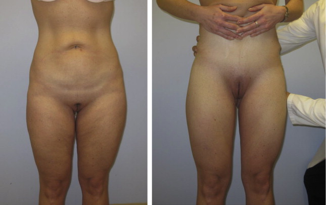
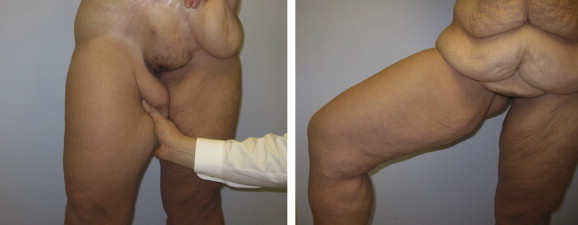
Physical examination should focus on a careful assessment of the medial thigh deformity and the variables outside the medial thighs potentially contributing to the deformity: soft tissue excess along the lower abdomen, mons pubis, hips, lateral thighs, and buttocks. In an effort to assess the variables contributing to the medial thigh deformity during the examination, patients should be asked to stand in front of a full-length mirror and to apply strong upward traction to the soft tissues of their lower abdomens with their 2 hands. At the same time, the surgeon or examiner should then apply strong upward traction to the patient’s lateral thighs and buttocks. This technique for examination helps eliminate the variables outside the medial thighs contributing to the medial thigh deformity. It also serves to show patients the function of circumferential body lifting procedures. Patients usually are not aware of the significance of these variables ( Fig. 3 ). A second maneuver that should also be considered is to have patients abduct one their thighs ( Fig. 4 ). Grasping the soft tissue of the medial thigh with the patient standing provides similar information. These techniques highlight soft tissue excess in the horizontal vector, accurately showing the function of the vertical medial thigh lift with liposuction procedure. The distal extent of the thigh deformity should be noted. Many women and some men have concerns about the excess soft tissue along the medial aspect of the knee and, in some instances, the leg as well ( Fig. 5 ). Some women following extreme weight loss have soft tissue draping the anterior aspect of the knee ( Fig. 6 ). Tissue elasticity can be assessed by evaluating the skin for the presence of striae and cellulite. The fat content of the thighs and surrounding structures should also be noted. Patients with less fat excess usually have a more deflated appearance (see Fig. 1 ). Any intertriginous dermatitis, potentially along the proximal medial thigh, is noted. Lymphedema and/or stigmata of lymphedema should be documented, as should the presence of arterial or vascular insufficiency. Scars from previous surgery along the lower body and extremities should be documented, as should contour irregularities secondary to liposuction. The quality of existing scars is important to note. Medial thigh lifting surgery may result in scars that are more perceptible than with other more traditional procedures.
Treatment goals and planned outcomes
A clear understanding of the variables contributing to a patient’s medial thigh deformity is critical to optimizing outcomes following medial thigh lift surgery. An analysis of the medial thighs is complicated by its dependent position. Unlike many other areas of the body commonly treated by plastic surgeons, the medial thigh contour is strongly affected by the status of the soft tissues immediately cephalad to it, particularly in patients after weight loss. A failure to address deformities along the mons pubis, hips, thighs and buttocks, or lower body produces suboptimal results. Circumferential lower body lift procedures effectively address these concerns (see Fig. 1 ). Efforts to approach the medial thighs before addressing the lower body deformities usually lead to undesirable outcomes ( Fig. 7 ). Equally important is the recognition of soft tissue excess in the horizontal vector. Almost all patients after weight loss and many individuals who have undergone significant liposuction have soft tissue excess in the horizontal vector.
Related posts:

Stay updated, free articles. Join our Telegram channel

Full access? Get Clinical Tree


