Groups no
Experimental groups
Purpose of testing
1.
Experimental
Nerve stump protected by epineural sheath jacket (ESJ)
To test the efficacy of ESJ application in prevention of neuroma formation
2.
Nerve stump protected by ESJ and buried into the adjacent muscle
To test if ESJ application combined with burying it into adjacent muscle is more effective than ESJ application alone
3.
Nerve stump protected by ESJ filled with autologous fat graft
To test if ESJ application combined with fat graft injection into ESJ is more effective than ESJ application alone
4.
Nerve stump protected by ESJ filled with autologous fat graft and buried into the adjacent muscle
To test if ESJ combined with fat graft injection into ESJ and burying it into adjacent muscle is more effective than ESJ application alone
5.
Control
Nerve stump left without any protection
To test the effect of neuroma formation
6.
Nerve stump buried into the adjacent muscle
To compare ESJ application with current standard of treatment in prevention of neuroma formation
Group 1: Nerve Stump Protected by Epineural Sheath Jacket (ESJ)
A 5 cm longitudinal incision was made on the right gluteal and thigh regions (Fig. 65.1). Gluteal muscles were cut and sciatic nerve was identified. Using blunt dissection, the plane between gluteus muscles and biceps femoris muscle was separated and the sciatic nerve was exposed. Dissection of sciatic nerve was performed from the arising point within pelvis bone structure to the bifurcation of common peroneal and tibial nerves (Fig. 65.2). The entire length of the exposed sciatic nerve was freed from surrounding tissue. Once whole sciatic nerve was dissected, a 2 cm long mid part of the sciatic nerve was resected creating a nerve defect (Fig. 65.3). When the transection of the nerve was made, fascicles started escaping due to natural endoneurial pressure and formed bulb-shaped formation at the proximal stump. Once the fascicles sprouting stopped, escaped fascicles were trimmed partially to avoid any fascicle interferences with applied ESJ. Harvested part of the sciatic nerve was suspended on a straight irrigator (30 ga) and all fascicles were removed using well- established pull out technique (Fig. 65.4). Thus an empty epineural sheath conduit was created. The distal part of the epineural sheath was trimmed creating a 7 mm long conduit. The lumen of the epineural sheath was slightly dilated using the jewelers microforceps. Thus the diameter of the epineural sheath was extended facilitating application of the epineural sheath over the proximal nerve stump and the conduit was threaded onto the proximal stump (Fig. 65.5, left column). Following this application, distal part of the epineural sheath was ligated using 10/0 nylon suture creating epineural sheath jacket. ESJ was attached over proximal stump of transected nerve using epineural sleeve technique. First two stitches were placed 180° apart on the medial or lateral margin of the proximal stump and grabbing margins of the ESJ. The highest attention should be paid not to catch any fascicle inside the nerve trunk. The sutures were placed proximally as much as possible over the proximal stump. Finally last two stitches between previously applied stay sutures were placed posteriorly and anteriorly. For Group 1–4 all steps of the surgical procedure were exactly the same as mentioned above. In Group 2–4 some additional steps were performed.
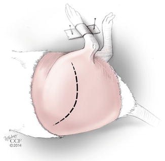
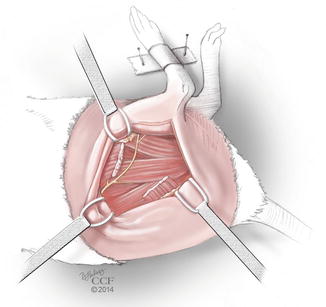
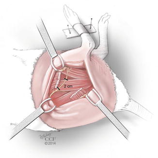
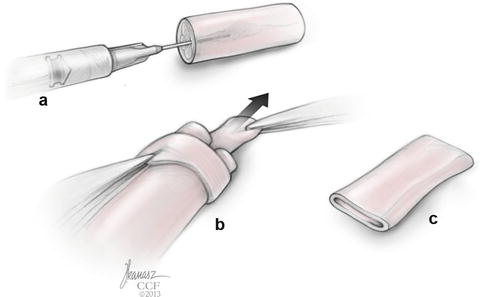
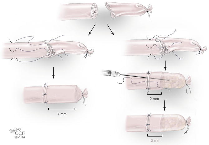

Fig. 65.1
Rat positioning and surgical incision

Fig. 65.2
Sciatic nerve exposure

Fig. 65.3
Nerve segment excision

Fig. 65.4
Nerve fascicles removal (a–c)

Fig. 65.5
Epineural Sheath Jacket application
Stay updated, free articles. Join our Telegram channel

Full access? Get Clinical Tree


