Introduction
The skull can be divided into two parts: the neurocranium, which forms a protective case around the brain, and the viscerocranium, which forms the skeleton of the face. This chapter details the viscerocranium and bones of the neurocranium that pertain to the viscerocranium.
Neurocranium
The neurocranium in adults is formed by a series of eight bones: the singular frontal, ethmoid, sphenoid, occipital bones centered on the midline, and the temporal and parietal bones occurring as bilateral pairs.1 The primarily flat frontal, parietal, and occipital bones form the calvaria (skullcap) by intramembranous ossification of head mesenchyme derived from the neural crest. The primarily irregular, yet considerably flat, sphenoid and temporal bones contribute to the cranial base via endochondral ossification of cartilage or from more than one type of ossification. The irregular ethmoid bone slightly contributes to the neurocranium but is primarily part of the viscerocranium. In reality, the flat bones and flat portions of the bones forming the neurocranium consist of convex external and concave internal curved surfaces.1
Fibrous interlocking sutures unite most calvarial bones in adulthood, although during childhood, the sphenoid and occipital bones are unified by synchondroses.2 Some sutures, comprising narrow closures of connective tissue at birth, remain open until adulthood. The sagittal suture is derived from neural crest cells and the coronal suture from paraxial mesoderm.2 The newborn skull contains fontanels, the most prominent being the anterior fontanel, which are widened sutures at points where more than two bones meet. The anterior fontanel, found where the two parietal and frontal bones meet, closes in most cases by 18 months of age, and the posterior fontanel closes by 1 to 2 months of age.2
Two primary centers of ossification traverse the frontal (metopic) suture in the second year, dividing the frontal bone into halves. Usually, the frontal suture disappears by age 6 years, when the halves fuse, but it can persist into adulthood as a metopic suture either totally, running from the midline of the glabella to the bregma, or partially.2 The glabella is a smooth anterior projecting prominence on the frontal bone superior to the root of the nose, and the bregma is the junction of the coronal and sagittal sutures.
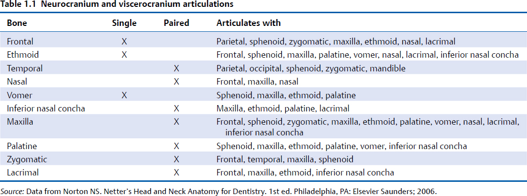
The maxillae and mandible provide the sockets and supporting bone for the maxillary and mandibular teeth. The maxillae contribute the greatest part of the upper facial skeleton, forming the skeleton of the upper jaw, which is fixed to the cranial base. (The mandible is detailed in Chapter 19.)
On the lateral aspect of the skull is the thin pterion. The pterion, located two finger breadths superior to the zygomatic arch and a thumb’s breadth posterior to the frontal process of the zygomatic bone, is formed by the articulations of the frontal, parietal, sphenoid, and temporal bones.1 The pterion overlies the anterior branch of the middle meningeal artery. Therefore, an injury to this region can damage the vessel, producing an epidural hematoma.1
The air-filled paranasal sinuses, including the maxillary, frontal, and ethmoidal sinuses, are discussed. The sphenoidal sinuses are discussed in Chapters 2 and 16. The bony articulations of the neurocranium and viscerocranium are described in Table 1.1, and the general processes of ossification are displayed in Table 1.2.
Frontal Bone
The frontal bone forms the forehead via its squamous, orbital, and nasal parts and two cavities, the frontal sinuses.
Squamous Part
The flat squamous part is the largest part of the frontal bone forming most of the forehead.3 The supraorbital margin of the frontal bone is the angular boundary between the squamous and the orbital parts (Fig. 1.1).4
On the external surface of the squamous part, about 3 cm above the midpoint of this margin, are the frontal tuberosities.4 These tubercles are more prominent in children and adult women. Ventrally, a shallow groove separates the frontal tuber-osities from the paired and curved superciliary arches.4 These arches extend laterally from the medially located, smooth, and elevated glabella and are more prominent in males. Partly dependent on frontal sinus size, supercilliary arch prominence is occasionally associated with small sinuses.4
The supraorbital notch (or foramen), which transmits the supraorbital vessels and nerve, lies at the junction between the sharp, lateral two-thirds and the rounded medial third of the supraorbital margin.4 The variably occurring frontal notch (or foramen) occurs medial to the supraorbital notch in 50% of skulls.4
Surgical Annotation
Recent interest in the surgical treatment of migraines has led to numerous anatomical studies identifying areas of nerve compression. The supraorbital nerve, as it emerges from the supraorbital foramen or notch, has been identified as a migraine trigger area.5 The supraorbital nerve can have compression from both a foramen as well as a notch as a result of the associated fascial bands. In addition to the soft tissue procedure, a supraorbital foraminotomy or fascial band release has been shown to improve postoperative outcomes.6 A transpalpebral incision can be used to access the supraorbital nerves to perform the decompression. An incision is made in the upper tarsal crease, with subsequent dissection identifying the supraorbital nerve. Muscles such as the corrugator supercilii are resected, and the foraminotomy is performed. Endoscopic techniques have also been described.7 With the use of the endoscopic technique, release of the zygomaticotemporal branch can also be performed.
The supraorbital margin extends laterally, forming the prominent zygomatic process, which articulates with the zygomatic bone. A posterosuperiorly curving line, which continues onto the squamous part of the temporal bone, divides into superior and inferior temporal lines.4 The temporal surface of the frontal bone is inferior and posterior relative to these temporal lines. The anterior surface of the temporal surface forms the anterior part of the temporal fossa. The rough inferior surface of the posterior margin of the squamous part articulates with the greater wing of the sphenoid.4
The nasal part of the frontal bone is discussed in the Nasal Bone: Nasal Bridge and Bony Septum section of this chapter. The interior surface of the frontal bone is detailed in Chapter 2.
Orbital Parts of the Frontal Bone
The two orbital parts of the frontal bone are thin, curved, and triangular laminae, consisting entirely of compact bone (Fig. 1.2).4 Forming the largest part of the orbital roofs, the orbital parts are separated by a wide, quadrilateral ethmoidal notch that is occupied by the cribriform plate of the ethmoid bone.4 The labyrinths of the ethmoid bone, which contain the ethmoidal air cells, articulate with the inferior surface of the lateral margins of the ethmoidal notch. This articulation converts two transverse grooves across each margin into anterior and posterior ethmoidal canals. These canals transmit the anterior and posterior ethmoidal nerves and vessels into the medial orbit.4
The posterolaterally ascending frontal sinuses open anterior to the ethmoidal notch and lateral to the nasal spine (Fig. 1.3). Deflecting from the median plane, these rarely symmetrical sinuses ascend between the frontal laminae and are separated by a thin septum.4 Each sinus communicates with the ipsilateral nasal cavity’s middle meatus via the frontonasal canal.4
Table 1.2 Neurocranium and viscerocranium ossification patterns
Bone | Parts | Ossification |
Frontal | Squamous, orbital, nasal portions | Intramembranous |
Ethmoid | Perpendicular plate, cribriform plate, ethmoid labyrinth | Endochondral |
Temporal | Squamous part, tympanic part | Intramembranous |
Nasal |
| Intramembranous |
Vomer |
| Intramembranous |
Inferior Nasal Concha |
| Endochondral |
Maxilla | Body; frontal, zygomatic, palatine, alveolar processes | Intramembranous |
Palatine | Perpendicular plate, horizontal plate, pyramidal process | Intramembranous |
Zygomatic | Frontal, temporal, maxillary processes | Intramembranous |
Lacrimal |
| Intramembranous |
Source: Data from Norton NS. Netter’s Head and Neck Anatomy for Dentistry. 1st ed. Philadelphia, PA: Elsevier Saunders; 2006.
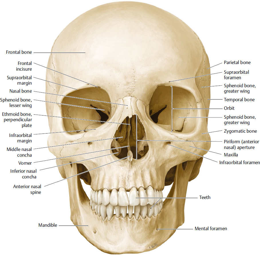
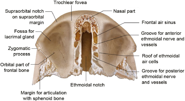
Fig. 1.2 Inferior view of the frontal bone. From this view, the ethmoidal notch and ethmoidal air sinuses can clearly be appreciated. Additional visibility of the orbital part surface features, including the fossa for the lacrimal gland, the sphenoidal articulating surface, and the zygomatic process, is obtained from this view.
Frontal Sinus Fractures
Surgical Annotation
Frontal sinus fractures can serve as a source of infection and cosmetic deformity. Approximately 10% of facial fractures involve the frontal sinuses.8 When outflow of the sinus is blocked as a result of injury to the nasofrontal duct, frontal sinus mucoceles can develop. In accessing the injury, management depends on which wall of the sinus is fractured, the extent of fracture displacement, and the involvement of the nasofrontal duct. Correction of the anterior table of the sinus, which is done mainly to correct the cosmetic deformity, can be performed through an existing laceration or coronal incision. When the posterior table is displaced, the coronal incision allows access to perform a cranialization, which involves removing the posterior wall and allowing for the sinus to be part of the intracranial cavity. The sinus mucosal surface must be removed and the outflow tract and dead space obliterated to prevent postinjury infection.
Different techniques for performing the obliteration have been described.8 Management of sinus preservation can also be performed with few complications depending on the fracture pattern.9
Each smooth and concave orbital surface contains a shallow anterolateral fossa for the lacrimal gland. The posterior border of the orbital plate articulates with the lesser wings of the sphenoid.4
Ethmoid Bone
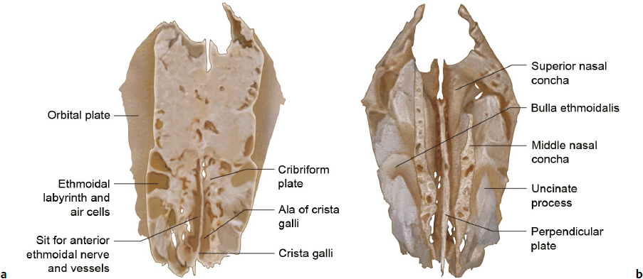
The fragile cuboidal ethmoid bone lies anteriorly in the cranial base, contributing to the medial orbital walls, nasal septum, roof, and lateral walls of the nasal cavity. The ethmoid is composed of a horizontal, perforated cribriform plate, a median perpendicular plate, and the two lateral labyrinths.3
Cribriform Plate
As mentioned, the horizontal cribriform plate fills the ethmoidal notch of the frontal bone (Fig. 1.3a). Penetrated by numerous foramina that contain olfactory nerve branches, the plate forms a large part of the nasal roof.3 The triangular and median crista galli projects superiorly from the plate and joins the frontal bone anteriorly via its two alae.3 Depressions in the cribriform plate on either side of the crista galli exist for the overlying olfactory bulb and gyrus rectus. Antero and lateral to the crista, foramina exist to transmit the anterior ethmoidal nerve and vessels from the nasal cavity to the foramen cecum.4
Perpendicular Plate
The perpendicular plate is discussed in the Nasal Bone: Nasal Bridge and Bony Septum section of this chapter.
Ethmoidal Labyrinths
On average, the ethmoidal labyrinths consist of 20 thin-walled air cells arranged as anterior, middle, and posterior groups containing 11, 3, and 6 air cells, respectively.4 The lateral orbital plate forms part of the medial orbital wall (Fig. 1.4). Adjoining articulations, save those that open into the nasal cavity, close all air cells. The air cells of the superior and posterior surfaces are closed by the ethmoidal notch of the frontal bone and both the sphenoidal conchae and orbital process of the palatine bone, respectively.4
The orbital plate covers the middle and posterior ethmoidal air cells articulating superiorly with the orbital plate of the frontal bone, anteriorly with the lacrimal bone, inferiorly with the maxilla and orbital process of the palatine bone, and posteriorly with the sphenoid bone.4 Anterior to the orbital plate, the lacrimal bone and frontal process of the maxilla complete the walls. Projecting posteroinferiorly from the labyrinth, the thin uncinate process crosses the ostium of the maxillary sinus to join the ethmoidal process of the inferior nasal concha.4
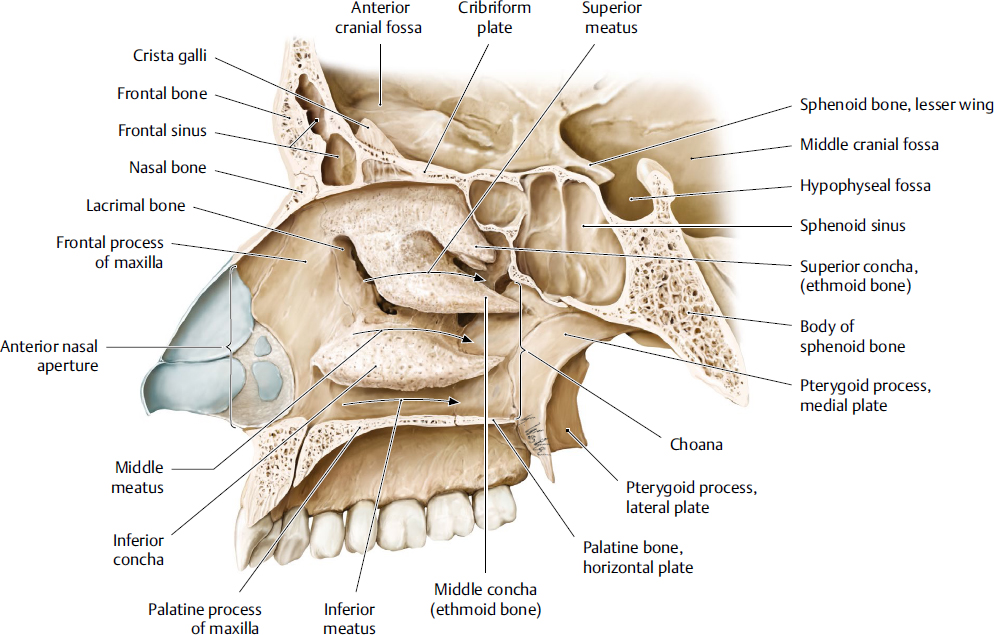
Fig. 1.4 Medial view of the right nasal cavity. This view of the nasal cavity allows better appreciation of the conchae, meatuses, portions of the hard palate, the frontal sinus, ethmoidal contributions to the nasal cavity, and the anterior nasal aperture. (Reproduced from THIEME Atlas of Anatomy, Head and Neuroanatomy, © Thieme 2010, Illustration by Karl Wesker.)
Descending from the inferior surface of the cribriform plate, the medial surface of the labyrinth forms part of the lateral nasal wall as the thin, lamellated, and convoluted middle nasal concha (Fig. 1.3b). Its anteroninferior lateral surface forms part of the middle meatus (Fig. 1.4).4 On the lateral wall of the middle meatus, middle ethmoidal air cells produce the ethmoidal bulla and open either on the bulla or above it. Posterior ethmoidal air cells open into the superior meatus, which is bounded by the superior nasal concha of the ethmoid.4 A curved infundibulum extends anteriorly and superiorly from the middle meatus, communicating with the anterior ethmoidal sinuses and in half of skulls continues superiorly as the frontonasal duct, which drains the frontal sinus.4
Temporal Bone
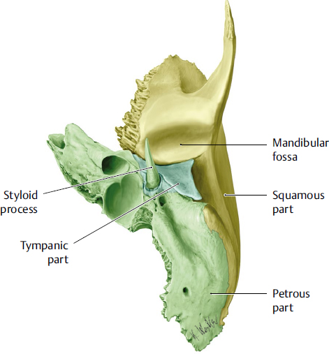
The paired temporal bones help form the base and lateral walls of the skull and are discussed in further detail in subsequent chapters (Fig. 1.1). The temporal bone houses the auditory and vestibular apparatuses and contains mastoid air cells.3 Each bone has eight centers of ossification that give rise to the three major centers observed before birth.2 The temporal bone comprises the squamous, petromastoid, and tympanic parts, as well as the styloid process.3 The temporal bone also has two associated canals. On its lateral surface, the external acoustic meatus conveys sound waves to the tympanic membrane. On its medial surface, the internal acoustic meatus conveys the facial and vestibulocochlear nerves.4
Squamous Part
The largest, squamous part lies anterosuperiorly and contains the thin and largest temporal portion, a zygomatic process and a mandibular fossa (Fig. 1.5).3 Its external temporal surface forms part of the temporal fossa. The concave, internal cerebral surface of the squamous part is grooved by the middle meningeal vessels.3 The lower border, fused to the anterior petrous part often contains traces of a petrosquamosal suture. Posteriorly, the squamous part fuses with the mastoid part. The anteroinferior border articulates with the greater wing of the sphenoid, and the superior border articulates with the inferior parietal bone at the squamosal suture.4
The zygomatic process extends anterolaterally from the squamous portion. It forms the zygomatic arch via an articulation between its obliquely posteroinferiorly sloping, deeply serrated anterior end, and the temporal process of the zygomatic bone.3 Its inferior surface forms a short articular tubercle, which contacts the articular disc of the temporomandibular joint and forms the anterior limit of the mandibular fossa.4
Related posts:


Stay updated, free articles. Join our Telegram channel

Full access? Get Clinical Tree


