Fig. 53.1
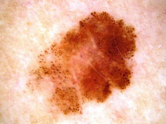
Nested melanoma of the elderly. A flat, irregularly pigmented and shaped lesion measuring 8 mm in a 65-year-old man
Dermatoscopic examination shows the presence of variably pigmented and irregularly distributed globules (Fig. 53.2), irregular blotches, and sometimes atypical pigment network, and confocal microscopy reveals the presence of a “cloud” pattern composed of large compact nests with variable atypia that is indicative of melanoma.


Fig. 53.2
Nested melanoma of the elderly. Dermatoscopic examination shows the presence of irregularly distributed globules
Pathology
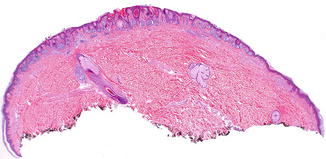
The architectural pattern is very similar to that of a junctional or superficial compound nevus (Fig. 53.3). The melanocytes are mostly grouped in distinct nests of the same size and shape and regularly distributed along the dermoepidermal junction (Fig. 53.4). There is variable cytologic atypia and only few, if any, melanocytes spreading throughout the epidermis associated with a lentiginous pattern (Fig. 53.5). The histological clues for diagnosis are the unusual findings of bizarre, large junctional nests in an old patient who should not have junctionally active nevi anymore and the presence of solar elastosis. FISH assay targeting RREB1, MYB, Cep6, and CCND1 has revealed chromosomal aberrations consistent with the standardized FISH diagnostic criteria for melanoma, and aCGH has identified multiple genomic aberrations.


