(1)
Department of Anatomy, Medical School Democritus University of Thrace, Alexandroupolis, Evros, Greece
Abstract
The cervical skin serves as an abundant source of flap material that can be transferred to close large facial defects. The superficial neck anatomic structures that are involved in skin flap surgery are the skin and the subcutaneous tissue, the platysma muscle, the investing layer of the deep cervical fascia, and the superficially located neck muscles. The skin of the neck is supplied by musculocutaneous and direct cutaneous perforators of branches of the external carotid artery and of the thyrocervical trunk of the subclavian artery. Important motor and sensory nerves that are located in a superficial level at the neck are involved in the surgery of the flaps that are derived from the neck. The regional neck flaps provide skin quite similar in color, texture, and thickness regarding the skin of the lateral face and must be the first choice when a local flap is not sufficient to reconstruct a sizeable defect.
The neck is the part of the body that joins the head and the trunk. It is bounded superiorly by the base of the cranium and the inferior border of the mandible and inferiorly by the thoracic inlet (Fig. 8.1).
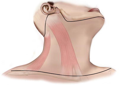

Fig. 8.1
Boundaries of the neck
More precisely, the superior limits run along the external occipital protuberance, the superior nuchal line, the mastoid apophyses, the anteroinferior borders of the external auditory canals, and the posterior and inferior mandibular borders. The lower limits lie along the superior border of the sternum and the clavicles, the acromioclavicular joints, and a line that joins the acromioclavicular joints to the spinous process of the seventh vertebra. A plane extending from the transverse vertebral processes to the anterior edges of the trapezius muscles divides the neck into a posterior and an anterior part.
8.1 Superficial Anatomy of the Anterior Neck
8.1.1 Skin and Subcutaneous Tissue
The skin that covers the neck although pliable is under tension due to the presence of the underlying platysma muscle. Its thickness is approximately 0.6 mm but becomes thicker at the submental area. It contains hair follicles, predominantly in the male, and sebaceous and sweat glands. During aging process, it becomes loose exhibiting a saggy appearance. Due to its pliability and mobility, greatly enhanced in the elderly, the skin of the neck constitutes an important tissue reservoir and is a first choice donor site for harvesting regional flaps nearly similar skin quality in the reconstruction of large facial defects.
The relaxed skin tension lines (RSTLs) at the neck run horizontally parallel one to each other (Fig. 8.2). Placing incisions transversely into wrinkles whenever possible, this hides the scars and enhances the cosmetic result. Τhe layer of subcutaneous tissue lies between the dermis of the skin and the deep cervical fascia. It contains various amounts of adipose tissue, cutaneous nerve filaments, and small blood and lymphatic vessels. The subcutaneous fat at the neck seems to form fat compartments in specific areas in a manner analogous to the superficial fat pads of the face. A fat compartment that is located at the submental region has been described in detail in recent studies (Hatef et al. 2009; Pilsl and Anderhuber 2010).
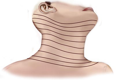

Fig. 8.2
Relaxed skin tension lines (RSTLs) at the neck
The platysma muscle (one of the superficial neck muscles; see below) is running within the subcutaneous tissue separating it into a thin superficial adipofascial layer that fixes the platysma tightly to the skin and a deep adipofascial layer consisting of loose fascial and poor fat tissue that enables the platysma to move over the underlying muscles (Imanishi et al. 2005). The subcutaneous dissection, when elevating a flap in the neck area that is covered by platysma muscle, has a dual form and is either subdermal or subplatysmal being demarcated by this muscle sheet.
All the anatomic structures are under the cover of the platysma. The subcutaneous tissue along with the platysma composes the superficial cervical fascia.
8.1.2 Platysma Muscle
The platysma is a superficial neck muscle located in its lateral aspect (Fig. 8.3); phylogenetically, it is accepted to be remnants of the panniculus carnosus. More precisely, it is a thin, broad, and quadrilateral muscle sheet and constitutes a major component of the SMAS layer. It is situated within the subcutaneous tissue and over the investing layer of the deep cervical fascia. Its length is usually about 20 cm and its width 10 cm. The platysma shows a great variability in size and thickness, may be absent or hypoplastic, and during aging process becomes thinner and difficult to determine.
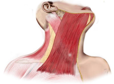

Fig. 8.3
The platysma muscle
The platysma originates from the superficial fascia over the upper part of the pectoralis major muscle in a plane corresponding even up to the 2nd and 3rd ribs and from the superficial fascia over the upper part of the deltoid muscle. The muscle fibers run parallel each other, upward, and medially crossing the clavicle toward the cheek. The medial border of the platysma ends at the submental region forming with its contralateral an inverted V, with the apex located in a variable level between the chin and the thyroid cartilage, either covering thus the submental area totally with muscle fibers or leaving it devoid of muscles fibers (de Castro 1980).
The lateral border of the platysma in the neck extends in a variable width beyond and over the external jugular vein. Entering the face, it continues in a curved fashion in a variable extent and ends at the labial commissure. After the fibers cross the mandible, they insert into the skin of the lower cheek and the oral commissure and blend with muscle fibers of the perioral muscles. It has been reported that the facial extent of the platysma may occupy more than 50 % of the face (Shah and Rosenberg 2009). At the mental area, some muscle fibers cross the midline and interlace with corresponding fibers of the contralateral muscle, and at the anterior part of the mandible, some others insert into the bone.
The platysma muscle receives its blood supply from a wide anastomotic network formed by arteries that distribute on each different part of the muscle. The upper part is supplied mainly by the submental artery and with contribution of direct perforators from the facial artery. The lower part is supplied by branches of the transverse cervical and the suprascapular artery, while the middle part by the superior thyroid artery. According to the posterior extent of the muscle, the occipital and the posterior auricular arteries may supplementary contribute to the vascularization of its lateral part. These arteries are described in detail below. According to its arterial supply, the platysma can be harvested as myocutaneous flap in three different types: having its pedicle superiorly, inferiorly, or laterally. The dominant arterial supply of the platysma comes from the submental artery and due to that fact a superiorly based platysma flap appears with enhanced vascularity and safety. The venous drainage of the platysma muscle is through the external jugular vein, the anterior jugular vein, the superior thyroid vein, the submental vein, and the facial vein (Uehara et al. 2001; Agarwal et al. 2004).
The platysma muscle is innervated by the cervical branch of the facial nerve which enters the muscle from its deep surface in the space between the mandibular angle and the sternocleidomastoid muscle. The marginal mandibular branches of the facial nerve run also deep to the platysma but do not take part in its innervation. The muscle is a lower lip and oral commissure depressor. When all of the muscle fibers contract, the skin of the neck wrinkles.
8.1.3 Investing Layer of the Deep Cervical Fascia
Beneath the platysma muscle lies the investing layer of the deep cervical fascia (Fig. 8.4). The investing layer of the deep cervical fascia is the outermost fascia of the three neck layers of the deep cervical fascia: the other two being the intermediate pretracheal layer and the deep prevertebral layer. It is a continuous sheet of fibrous tissue, encircling completely the neck. It attaches posteriorly to the spines of the cervical vertebrae and to the nuchal ligament. Passing anteriorly around the neck, it splits and envelopes first the trapezius muscle and then the sternocleidomastoid muscle and the submandibular gland. The investing layer forms the roof of the posterior triangle of the neck and the floor of the submandibular triangle. In the midline it is attached to the chin, the body of the hyoid bone and the manubrium of the sternum. Inferiorly, the investing layer is attached to the sternum, the clavicle, and the acromion. Superiorly, it is attached to the external occipital protuberance, the superior nuchal lines, the tip of the mastoid process, the tympanic plate, and the styloid process. It splits at the lower border of the mandible into a medial and a lateral layer which continue up to the cheek.
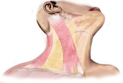

Fig. 8.4
The investing layer of the deep cervical fascia
The pretracheal layer is found below the hyoid bone; surrounds the trachea, the esophagus, the larynx, and the thyroid gland; and envelopes the infrahyoid strap muscles.
The prevertebral layer is the innermost cervical fascia and invests the prevertebral muscles and encloses the scalene and the levator scapulae muscles. The investing layer of the deep cervical fascia is the fascia commonly associated with flap surgery. Likewise, the fascial layers of the head are the source of considerable controversy with respect to the organization and nomenclature of the investing layer of the neck, while some authors even challenge its existence (Zhang and Lee 2002; Nash et al. 2005).
8.1.4 Sternocleidomastoid Muscle
Apart from the platysma muscle, the sternocleidomastoid and the anterior borders of the trapezius muscles are encountered in a superficial level just beneath the platysma level (Fig. 8.5).
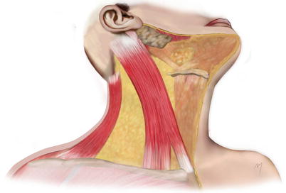

Fig. 8.5
The sternocleidomastoid muscle and the trapezoid muscle at the neck
The sternocleidomastoid muscle is originated by two heads, a medial termed the sternal head and a lateral one termed the clavicular head. The sternal head originates from the upper part of the anterior surface of the manubrium sterni and runs superolateral and posterior in a slightly oblique direction. The clavicular head originates from the upper surface of the medial third of the clavicle and runs upward in a more vertical direction than the sternal head. There are cases where the clavicular head exhibits variability in its origin breadth, being as narrow as the sternal head or widening to the origin of the trapezius.
The sternal and the clavicular heads as they run upward merge together in a level between the middle and the lower third of the neck and form a thick and round muscle belly. The muscle continues to the mastoid region passing obliquely to the side of the neck. It inserts by a tendon into the lateral surface of the mastoid process and, by an aponeurosis, into the lateral half of the superior nuchal line.
The upper part of the muscle is supplied from branches of the occipital and small contribution of branches of the posterior auricular arteries in its most upper part. Despite this, the sternocleidomastoid branch of the occipital artery has also been reported to supply lower levels of the sternocleidomastoid muscle (Fróes et al. 1999). Its middle part is supplied by the sternocleidomastoid branch of the superior thyroid artery. The lower part of the muscle is supplied in more than 80 % by a branch arising from the suprascapular artery, and in the cases where this branch does not exist, a longer sternocleidomastoid branch of the superior thyroid artery or branches from the transverse or superficial cervical arteries undertake the blood supply (Kierner et al. 1999).
The sternocleidomastoid muscle is innervated by the accessory nerve.
When the muscle acts unilateral, it tilts the head to its own side and simultaneously rotates it so as turning the face toward the opposite side. When acting bilateral, it draws the head forward.
8.1.5 Trapezius Muscle
The trapezius muscle (see Chap. 2) is also a muscle of the upper limb. Its upper part and especially its anterior margin constitute the most posteriorly located superficial muscle of the lateral neck (Fig. 8.5). The anterior margin of the trapezius makes up also the posterior border of the posterior neck triangle and is a landmark in the design of flaps (e.g., cervicopectoral flap). Even though the anterior muscle margin is easily palpated, this becomes somewhat difficult in the anesthetized patient where the muscle is under relaxation.
8.1.6 Triangles of the Neck
The side of the neck is divided by the sternocleidomastoid muscle, which crosses the neck in an oblique direction, into an anterior and a posterior triangle (Fig. 8.6).
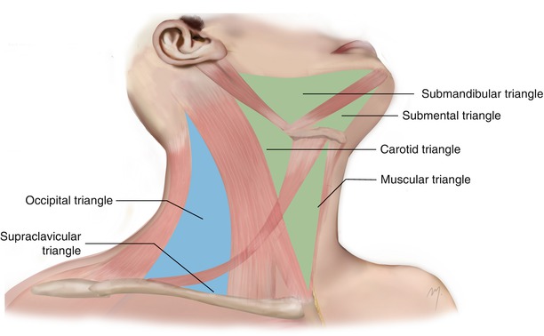

Fig. 8.6
The triangles of the neck. Blue color indicates the posterior triangle and the green color the anterior triangle
8.1.6.1 Anterior Triangle
The base of the anterior triangle lies superiorly and is formed by the border of the mandible and a line that extends from the mandibular angle to the mastoid process, while its apex is situated inferior at the sternum. Anteriorly, it is bounded by the midline of the neck and posteriorly by the anterior margin of the sternocleidomastoid.
The anterior triangle is further subdivided into the following:
8.1.6.1.1 Submental Triangle
The submental triangle has its base at the body of the hyoid bone and its apex at the chin. The anterior bellies of both digastric muscles form the side pleura of the triangle. The mylohyoid muscles form its floor. The contents of the triangle are the submental lymph nodes, some small veins that unite to form the anterior jugular vein, and the terminal part of the submental artery as it turns to ascend to the chin.
8.1.6.1.2 Submandibular Triangle
The submandibular triangle (digastric triangle) is bounded superiorly by the border of the mandible and the line extending from its angle to the mastoid process, posteriorly by the posterior belly of the digastric and the stylohyoid muscle, and anteriorly by the anterior belly of the digastric muscle. The mylohyoid, hyoglossus, and constrictor pharyngis muscles form the floor of the submandibular triangle.
The structures that are of interest in flap surgery when dissecting in a subplatysmal plane in the area of the submandibular triangle are the facial vein, the facial artery as it curves around the mandibular border to enter the face, the submental artery, and the cervical and marginal branches of the facial nerve.
8.1.6.1.3 Carotid Triangle
The boundaries of the carotid triangle are superiorly the posterior belly of the digastric and the stylohyoid muscles, anteriorly the superior belly of the omohyoid muscle, and posteriorly the anterior border of the sternocleidomastoid muscle. The inferior and middle pharyngeal muscles form the floor of the triangle. When removing the platysma, a part of the external and the anterior jugular veins and cutaneous cervical branches appear in the superficial level of the triangle.
8.1.6.1.4 Muscular Triangle
The borders of the muscular or visceral triangle of the neck are the midline of the neck anteriorly, the superior belly of omohyoid muscle superiorly, and the anterior margin of the sternocleidomastoid inferiorly. It contains the muscles of the neck: sternohyoid, superior belly of omohyoid, sternothyroid, and thyrohyoid. The first two form the superficial layer of these muscles.
8.1.6.2 Posterior Triangle
The base of the posterior triangle corresponds to the superior surface of the middle third of the clavicle and its apex at the superior nuchal line at the point where the sternocleidomastoid and trapezius muscles approximate to each other. Anteriorly, the posterior triangle is bordered by the posterior border of sternocleidomastoid and posteriorly by the anterior margin of the trapezius. The posterior triangle is subdivided into the occipital and the supraclavicular triangles by the inferior belly of the omohyoid muscle, which runs crossing the space about 2–2.5 cm above the clavicle.
8.1.6.2.1 Occipital Triangle
The occipital triangle is the upper and larger part of the posterior triangle. Its floor is formed from the top down by the semispinalis capitis at the apex, the splenius capitis, the lavatory scapulae, the scalenus posterior, and the scalenus medius muscles.
The platysma covers the lower part of the triangle in a various degree.
The important surface structures found at this triangle are the branches of the cervical plexus as they radiate emerging from the posterior border of the sternocleidomastoid muscle and the spinal accessory nerve as it emerges slightly superiorly and runs obliquely, crossing the triangle in its middle to reach the trapezius. All of these neural branches emerge from the posterior border of the sternocleidomastoid within 2 cm above or below a point termed Erb’s point. Erb’s point is located at the posterior border of the sternocleidomastoid muscle approximately in the mid of its mastoid and sternoclavicular attachments and serves as an important landmark. Identifying this point is useful not only in locating the emerging point of the mentioned nerves so as to protect them during surgery but also in blocking the cutaneous nerves of the cervical plexus by gaining anesthesia of almost the bigger part of the neck skin.
8.1.6.2.2 Supraclavicular Triangle
The supraclavicular triangle (omoclavicular triangle) is the lower and smaller part of the posterior triangle with a variable size that depends on the outspread of the clavicular attachments of the sternocleidomastoid and trapezius muscles. The first rib, the scalenus medius, and a part of the scalenus anterior and the first slip of the serratus anterior muscle form its floor. The triangle is covered almost fully by the platysma and in the superficial level is crossed by the supraclavicular nerves and the external jugular vein as it descends across the posterior edge of the sternocleidomastoid.
8.1.7 Arteries That Supply the Skin of the Neck
The skin of the neck is supplied by musculocutaneous and direct cutaneous perforators of branches of the external carotid artery and of the thyrocervical trunk of the subclavian artery.
8.1.7.1 Branches of the External Carotid Artery That Contribute to the Neck Skin Supply
8.1.7.1.1 Facial Artery (Cervical Part)
The facial artery arises from the external carotid artery, as one of its anterior branches, at the carotid triangle (Fig. 8.7). It usually ends at the inner canthus as angular artery and anastomoses with the dorsal nasal artery (see Chap. 4). For descriptive purposes, this long course is divided in a cervical and a facial part according to the area where the vessels run.
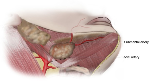

Fig. 8.7
The cervical part of the facial artery
The facial artery arises as an anterior branch of the carotid artery immediately above the greater horn of the hyoid bone and above the lingual artery (Fig. 8.7). It lies beneath the platysma and the hypoglossal nerve often runs above it. Its diameter ranges from 1.7 to 3.6 mm (mean 2.6 mm) (Zhao et al. 2000). In about 20 % of the cases, the facial and the lingual arteries arise with a common trunk from the external carotid artery (Shima et al. 1998).
The facial artery after its origin runs upward and forward, deep to the stylohyoid and the posterior belly of the digastric. Above the stylohyoid, it turns down and forward between the medial pterygoid muscle and the posterior aspect of the submandibular gland. As it reaches the lower border of the mandible, it curves around it, just in front of the anterior edge of the masseter muscle; pierces the deep fascia, lying immediately deep to platysma; and enters the face, continuing as the facial part, running toward the alar base, and giving off its branches to the face (see Chap. 5). At the point where the artery crosses the mandible, it lies very superficial, and its pulsation is most palpable. The facial artery, with its accompanying vein, crosses deep the mandibular branch of the facial nerve.
In the neck, the facial artery gives off the ascending palatine, the tonsillar, the glandular, and the submental arteries. The ascending palatine artery arises close to the origin of the facial artery, runs up between styloglossus and stylopharyngeus to the side of the pharynx, and ascends along it, between the superior constrictor of the pharynx and the medial pterygoid toward the skull base. Its branches supply the soft palate, the tonsils, and the auditory tube and anastomose with its opposite fellow, the greater palatine branch of the maxillary artery and the tonsillar and the ascending pharyngeal arteries.
The tonsillar artery ascends between medial pterygoid and styloglossus and after penetrating the superior constrictor of the pharynx enters the tonsil. It supplies the tonsil and the root of the tongue. The glandular branches are 3–4 and supply the submandibular salivary gland, lymph nodes, and a small area of overlying skin as cutaneous perforators.
8.1.7.1.2 Submental Artery
The submental artery constitutes the largest cervical branch of the facial artery. It branches off at a point deep to the submandibular gland or at its superior edge as the facial artery separates from the submandibular gland. The artery originates in a point of 5–7 mm from the border of the mandible (Martin et al. 1993; Magden et al. 2004). At its origin, the mean diameter of the artery has been reported ranging between 1 and 2 mm (Martin et al. 1993; Faltaous and Yetman 1996; Pinar et al. 2005a, b).
It runs forward on the surface of the mylohyoid below the border of the mandibular body. As it reaches the anterior belly of the digastric muscle, it continues running either deep to it in 70 % or superficial to it in 30 % of the cases (Faltaous and Yetman 1996). At the symphysis of the mandible, it turns around the lower border of the mandible body, enters the chin, and divides into a superficial branch and deep branch, which anastomose with the inferior labial and mental arteries, supplying the chin and the lower lip (see Chap. 6).
During its course in the submandibular triangle, the artery gives off numerous small branches to the submandibular and sublingual glands and to the adjacent muscles and platysma. It also gives off perforating cutaneous branches in a number of 1–4 (Curran et al. 1997; Kim et al. 2002a, b) (Fig. 8.8). These branches pierce the platysma, supply the skin over the submental triangle, and anastomose with their contralaterals, providing a strong arterial pedicle for designing flaps in the submental region. Moreover, the submental artery anastomoses with the mental, the inferior labial, and the sublingual arteries, a network that is of great importance to the blood supply of platysma muscle and the superiorly based platysma flap (Lasjaunias et al. 1979).
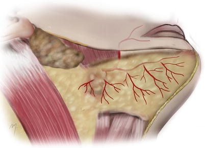

Fig. 8.8
Submental artery cutaneous perforators
8.1.7.1.3 Superior Thyroid Artery
The superior thyroid artery is described as the first branch of the external carotid artery. It arises beneath the anterior border of the sternocleidomastoid muscle just below the level of the greater horn of the hyoid bone (Fig. 8.9). In relation to the carotid bifurcation, it arises in 65 % of the cases at the same level or above it. In 35 % of the cases, it arises below the level of bifurcation from the common carotid artery (Ozgur et al. 2009).
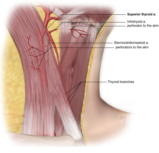

Fig. 8.9
Superior thyroid artery
It courses inferiorly and medially, to the apex of the lobe of the thyroid gland. In its course in the carotid triangle, the superior thyroid artery lies only beneath the skin and the platysma. As it descends, it then passes beneath the omohyoid and sternothyroid muscles with the external laryngeal nerve usually lying medially to it. The superior thyroid artery ends at the thyroid gland where it divides into the anterior, isthmal, posterior, and lateral thyroid final branches.
Through its course, the superior thyroid artery gives off the following branches: the infrahyoid artery, the superior laryngeal artery, the sternocleidomastoid artery, the cricothyroid artery, and the final thyroid branches. The branches of the superior thyroid artery that contribute to the neck skin supply are the infrahyoid and the sternocleidomastoid arteries.
8.1.7.1.4 Infrahyoid Artery
This is a small branch running along the lower border of the hyoid bone and supplies the superior portion of the infrahyoid strap muscles and the overlying part of the skin.
8.1.7.1.5 Sternocleidomastoid Artery
The sternocleidomastoid branch arises from the superior thyroid artery in about 80–85 % of the cases, while in the remainder it originates directly from the external carotid artery (Hu et al. 2006; Ozgur et al. 2009). When it originates from the superior thyroid artery, this occurs approximately 0.5–1 cm from the origin of the superior thyroid artery (Hurwitz et al. 1983). It courses downward and laterally across the carotid sheath and at the medial margin of the middle portion of the sternocleidomastoid muscle enters the muscle on its deep surface. The precise point where the sternocleidomastoid artery enters the muscle is situated in a distance of 3 cm away from the origin of the superior thyroid artery (Hu et al. 2006). Small perforators perforate the mid-third of the sternocleidomastoid muscle and supply the overlying platysma and skin. In the same area, a mostly constant direct cutaneous perforator has been found to emerge at the anterior border of the sternocleidomastoid muscle in its midportion (Fig. 8.10). This branch travels initially below the platysma, and after piercing it courses subcutaneously to the midline adjoining its contralateral supplying a large area of the anterior neck skin (Hurwitz et al. 1983; Wilson et al. 2012). Recently, Wilson et al. (2012) raised long perforator flaps that extended even beyond the midline, pedicled on just one of these cutaneous perforators.
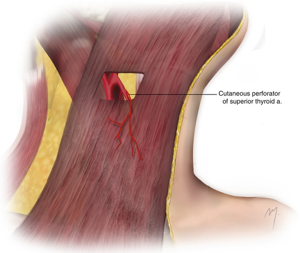

Fig. 8.10
Cutaneous perforator of the superior thyroid artery
The infrahyoid artery anastomoses with its contralateral in the midline. Anastomoses between the muscular branches of the superior thyroid artery and the muscular branches of the inferior thyroid artery (which supplies the inferior portion of these muscles) are formed passing through the entire musculature (Eliachar et al. 1984).
8.1.7.1.6 Occipital Artery: Sternocleidomastoid Branches
The occipital artery (see Chap. 2) contributes to the vascular supply of the upper part of the neck by two small branches, the lower and the upper sternocleidomastoid branches (Fig. 8.11).
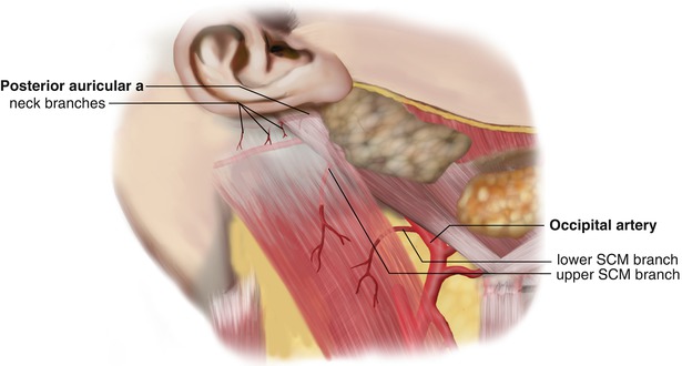

Fig. 8.11
Musculocutaneous perforators of the lower and upper sternocleidomastoid branches of the occipital artery and musculocutaneous perforators of the posterior auricular artery
The occipital artery branches from the posterior aspect of the external carotid artery about 2 cm from its origin and opposite to the origin of the facial artery. It courses up and back deep to the posterior belly of the digastric muscle and in front of the internal carotid artery, the internal jugular vein, and the vagus, the hypoglossal, and the accessory nerves. Running to the occipital area, it passes in the occipital groove and the medial aspect of the mastoid process deep to the attachments of the sternocleidomastoid muscle. At this part, it gives off the lower and the upper sternocleidomastoid branches.
The lower sternocleidomastoid branch arises from the starting point of the occipital artery, courses posteroinferiorly over the hypoglossal nerve, and enters the sternocleidomastoid muscle. The upper sternocleidomastoid branch arises from the occipital artery, after a short distance from the previous one, immediately after it crosses the accessory nerve. This branch runs in a same posteroinferiorly course in company with the accessory nerve entering the deep surface of the sternocleidomastoid muscle. Musculocutaneous perforators of both of the branches supply the upper third of the sternocleidomastoid muscle and a quite large area of the upper neck skin.
8.1.7.1.7 Posterior Auricular Artery: Neck Branches
The posterior auricular artery supplies the digastric and the stylohyoid muscles as well as the parotid gland. The artery gives off small branches to the neck that contribute to the vascularization of the uppermost part of the sternocleidomastoid and the overlying skin (Fig. 8.11).
8.1.7.2 Branches of the Thyrocervical Trunk Contributing to the Neck Skin Supply
8.1.7.2.1 Inferior Thyroid Artery
The inferior thyroid artery is usually branched from the thyrocervical trunk of the subclavian artery (Fig. 8.12). It ascends anterior to the medial border of the scalenus anterior muscle. At the level below the sixth cervical transverse process, it turns medially situated between the vertebral vessels and the carotid sheath. Finally, it descends to the lower border of the thyroid gland, lying on the longus colli muscle and coming in a variable relation to the recurrent laryngeal nerve (Tang et al. 2012).
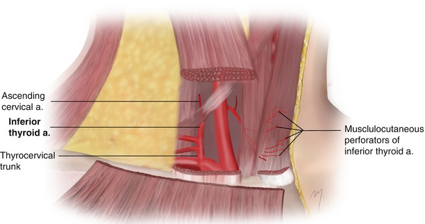

Fig. 8.12
The inferior thyroid artery and its branches. Perforators through the muscular branches supply the skin
The inferior thyroid artery gives off the inferior laryngeal artery, the ascending cervical artery, and pharyngeal, tracheal, esophageal, thyroid, and muscular branches. The inferior laryngeal artery runs upward on the trachea accompanied by the recurrent laryngeal nerve and enters the larynx supplying the laryngeal muscles and its mucous membrane. The ascending cervical artery arises at the point where the inferior thyroid artery turns medially to run behind the carotid sheath. It runs upward on the anterior tubercles of the transverse processes of the cervical vertebrae situated between the scalenus anterior and longus capitis muscles. It supplies the neighboring neck muscles and gives off spinal branches into the vertebral canal. The artery anastomoses with the deep cervical, occipital, and ascending pharyngeal arteries.
The pharyngeal, tracheal, and esophageal branches supply the lower part of the pharynx, the trachea, and the esophagus. The thyroid branches supply the thyroid and parathyroid glands and anastomose with the superior thyroid artery glandular branches.
8.1.7.2.2 Muscular Branches
Stay updated, free articles. Join our Telegram channel

Full access? Get Clinical Tree


