Introduction
The nasal cavity is located in the central region of the face and is divided by the nasal septum into a pair of cavities. These two cavities constitute the uppermost portion of the respiratory tract and continue anteriorly to the outer environment via the nares and posteriorly to the nasopharynx via the choanae. The nasal cavity is divided into two regions: the nasal vestibule and the nasal cavity. The nasal vestibule is the initial part of the nasal cavity as entered through the nares; it is lined by epithelium and contains hair (vibrissae) and sebaceous glands. The nasal cavity is the large space following the nasal vestibule and is lined by mucosa. It is divided into four meatuses (the superior, middle, inferior, and common meatus) by three nasal conchae that arise medially from the lateral nasal wall and curve inferiorly. Each meatus with the exception of the common meatus is located inferior to the conchae. The superior meatus is inferior to the superior concha, the middle meatus is inferior to the middle concha, and the inferior meatus is inferior to the inferior concha. The common meatus is the medial portion of the nasal cavity; it is located on both sides of the nasal septum and extends vertically. The nasal cavity is also divided into two regions according to function: the olfactory region and the respiratory region. The olfactory region is located in the upper part of the nasal cavity and is lined by olfactory epithelium, which contains olfactory receptors. The residual part of the nasal cavity has a respiratory function.1–3
Roof of the Nasal Cavity
The roof of the nasal cavity comprises the nasal bone, frontal bone, ethmoid bone, and sphenoid bone (Fig. 16.1). In the coronal section, it is triangular; the roof is extremely narrow, and the floor is wide. In a sagittal section, its height is greatest in the central region, which contains the cribriform plate of the ethmoid bone; the roof then decreases in height as it progresses in the anterior and posterior directions. The roof is adjacent to three paranasal sinuses: the frontal sinus, sphenoid sinus, and ethmoid sinus.1–3
Floor of the Nasal Cavity
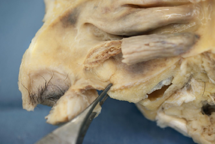
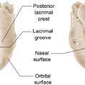
The floor of the nasal cavity mainly consists of the superior surface of the maxilla and palatine bone, which together constitute the hard palate (Fig. 16.2). The maxilla occupies the anterior two-thirds of the floor, and the palatine bone occupies the posterior one-third. The incisive canal is located in the anterior part of the floor, immediately lateral to the nasal septum. The nasopalatine nerve and terminal end of the greater palatine artery pass through the incisive canal.1–3
Medial Wall (Nasal Septum)
The nasal septum forms the medial wall of the nasal cavity and divides the entire nasal cavity into two (Fig. 16.3). Histologically, the nasal septum is made up of a cartilaginous part and a bony part. The septal nasal cartilage forms the cartilaginous part and occupies the anterior part of the nasal septum. The posterior part is the bony part and is made up primarily of the vomer in the inferior posterior region and the perpendicular plate of the ethmoid bone in superior region. The most antero-inferior part of the nasal septum, which is positioned more anteroinferior to the edge of the nasal septal cartilage, is called the columella. The nasal septum often curves and shifts to either the right or left and sometimes obstructs the common nasal meatus on one side. The reported frequency of nontraumatic septal deviation is 20% to 58%.4–6 Guyuron et al7 classified septonasal deviation into six types: septal tilt, C-shaped deformity (either anteroposterior or cephalocaudal), S-shaped deformity (either anteroposterior or cephalocaudal), and localized deformity. The most common type of deviation is the septal tilt type, in which the septum has no curve but is tilted toward one side in the coronal section. This type is observed in approximately 40% of patients with septonasal deviation. The second most common type is the C-shaped anteroposterior deviation, which occurs in approximately 32% of patients. In this type, the septum exhibits a C-shaped curve in the coronal section. The C-shaped cephalocaudal deviation, in which the septum forms a C shape in the coronal section, is observed in approximately 4% of patients. Anteroposterior and cephalocaudal S-shaped deviations are observed in 9% and 1% of patients, respectively. Finally, localized deformities are observed in about 14% of patients with septonasal deviation.7
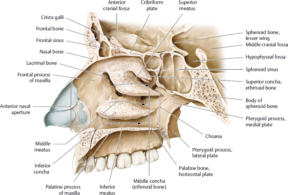
Fig. 16.1 Sagittal section of the nasal cavity (right side). (From THIEME Atlas of Anatomy, Head and Neuroanatomy. © Thieme 2010, Illustration by Karl Wesker.)
An important region of the nasal septum is Kiesselbach’s or Little’s area, located in the anterior part of the nasal septum. Five arteries supplying the nasal septum anastomose at this point to create an arterial plexus (Fig. 16.4). This area is frequently involved in chronic epistaxis, especially in children.
Lateral Wall of the Nasal Cavity
The lateral wall of the nasal cavity is characterized by three conchae that are long in the anteroposterior direction and protrude medially toward the nasal cavity; their free edge rolls inferiorly (Fig. 16.4). These conchae are termed the superior nasal concha, middle nasal concha, and inferior nasal concha, and the meatuses below these conchae are called the superior nasal meatus, middle nasal meatus, and inferior nasal meatus, respectively. At the posterior end of these conchae, the three meatuses conflate and meet at the nasopharynx; this point is termed the nasopharyngeal meatus. An atrophic supreme nasal concha is sometimes found above the superior concha.8 The posterosuperior part of the nasal cavity, which forms the space between the nasal roof and superior concha, is the sphenoethmoidal recess.
Some important structures are located around the middle meatus. The ethmoid bulla, which contains the ethmoid bulla cells of the ethmoid sinus, projects from the medial wall of the orbit. Part of the ethmoid bulla is exposed on the superior part of the lateral wall of the middle nasal meatus. The uncinate process is the thin, bony projection located above the attachment of the inferior nasal concha. The deep groove between the ethmoid bulla and uncinate process is called the semilunar hiatus. The semilunar hiatus is divided into two parts in its anterior region by the projection of the ethmoid bulla: the superior and inferior semilunar hiatus. These structures are involved in connecting the paranasal sinuses. The ethmoidal infundibulum is the funnel-shaped space inside the medial wall of the nasal cavity. It is surrounded by the uncinate process medially and ethmoid bulla laterally and continues to the semilunar hiatus posteriorly. The bony bulge anterior to the anterior end of the middle nasal concha is called the agger nasi. The outward bulge immediately anterior to the middle meatus is called the antrum of the middle meatus.1–3
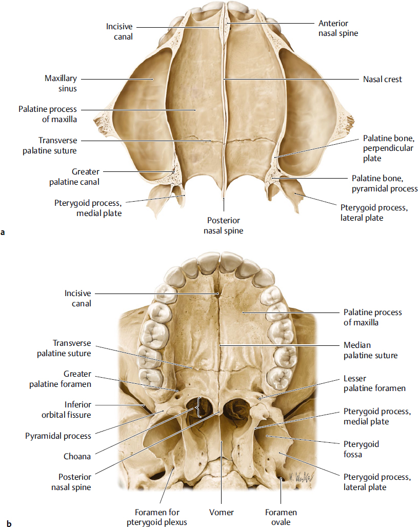
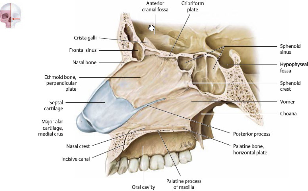
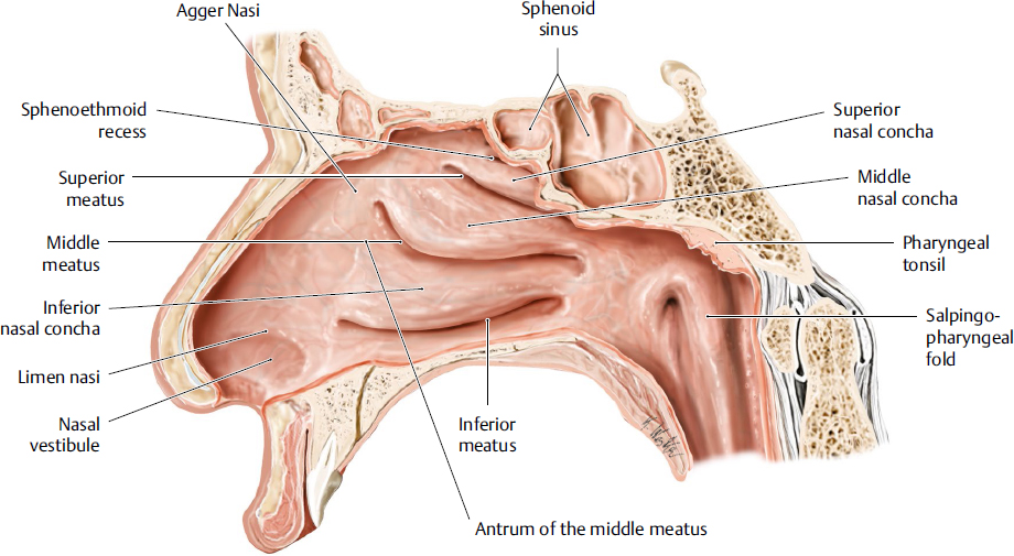
Fig. 16.4 Right lateral wall of the nasal cavity. (Modified from THIEME Atlas of Anatomy, Head and Neuroanatomy. © Thieme, 2010, Illustration by Karl Wesker.)
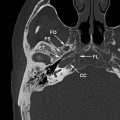
The opening of the nasolacrimal duct is located in the antero-superior part of the inferior meatus, just below the attachment of the inferior concha (Fig. 16.5). The openings of each paranasal sinus and the nasolacrimal duct are described in the section that discusses the paranasal sinuses, nasolacrimal ducts, and their openings into the nasal cavity.
Blood Supply of the Nasal Cavity
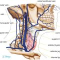
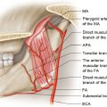
The nasal cavity, including both the medial and lateral walls, is supplied by five arteries: the anterior ethmoidal artery, posterior ethmoidal artery, sphenopalatine artery, greater palatine artery, and superior labial artery (Fig. 16.6).2
Both the anterior ethmoidal artery and posterior ethmoidal artery are branches of the ophthalmic artery. They branch within the orbital cavity, enter the nasal cavity through the ethmoidal bone, and mainly supply the upper region of the cavity. A branch of the anterior ethmoidal artery runs anteriorly, pierces the nasal bone, and distributes its blood supply to the nasal epithelium. The main artery supplying the nasal cavity is the sphenopalatine artery. This artery arises from the maxillary artery in the pterygopalatine fossa and enters the nasal cavity through the sphenopalatine foramen, which is located on the lateral wall posterior to the superior nasal meatus. After entering the nasal cavity, the artery gives off various branches. The two main branches course to the posterior part of the nasal septum and posterior part of the lateral nasal wall. The greater palatine artery, which is the terminal branch of the descending palatine artery, enters the nasal cavity through the incisive canal and distributes its blood supply to the lower part of the nasal cavity. The superior labial artery, which is a branch of the facial artery, also enters the nasal cavity through the soft tissue in the upper lip and distributes its blood supply to the anterior and lower parts of the nasal cavity. These arteries anastomose with one another on both the lateral nasal wall and the nasal septum. The region in which these five arteries meet and anastomose at the anterior part of the nasal septum is called Kiesselbach’s area (Fig. 16.6b).1–3,9
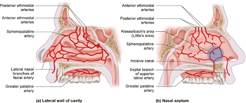
Fig. 16.6 Arterial blood supply of the nasal cavity. (a) Arterial blood supply of the nasal septum. Kiesselbach’s area is located in the anterior part of the nasal septum. Five arteries supplying the nasal septum anastomose in this region and form the arterial plexus. (b) Arterial blood supply of the lateral nasal wall.
Sensory Innervation of the Nasal Cavity
The sensory innervation of the nasal cavity is separated into two areas by the line connecting the anterior nasal spine and the sphenoethmoidal recess (Fig. 16.7). The upper area of the nasal cavity contains the anterior ethmoidal nerve, which is a branch of the ophthalmic nerve (first division of the trigeminal nerve). This nerve, accompanied by the anterior ethmoidal artery, enters the nasal cavity through the ethmoid bone and courses through the anterior and superior parts of the nasal cavity. The lower area of the nasal cavity is innervated by a branch derived from the maxillary nerve (second division of the trigeminal nerve). The nasopalatine nerve and medial superior posterior branch of the greater palatine nerve innervate the nasal septum in this region of the nasal cavity. The medial superior posterior branch innervates the upper half of this area, and the nasopalatine nerve innervates the lower half. The lateral superior posterior nasal branch and inferior posterior nasal branch of the greater palatine nerve innervate the lateral nasal wall in this region of the nasal cavity.

Related posts:
Stay updated, free articles. Join our Telegram channel

Full access? Get Clinical Tree


