Fig. 30.1
Myxofibrosarcoma presenting with a gradually enlarging painless mass with erythema and a tendency for diffusely infiltrative growth on the leg
Pathology
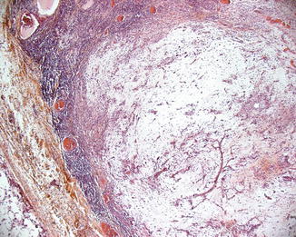
MFS presenting in the skin involves the reticular dermis, although the main bulk of the tumor is located in the subcutaneous tissue (Fig. 30.1). Superficial portions of MFS often appear benign, while deeper regions contain more obvious histomorphologic evidence of malignancy. Consequently, superficial cutaneous biopsies may result in an underestimation of tumor grade or even misdiagnosis as a benign process.
MFS is usually multinodular with ill-defined, deceptively infiltrative margins (macroscopically and microscopically) that extend into surrounding tissues, resulting in distant microscopic tumor deposits that predispose to local recurrence after resection.
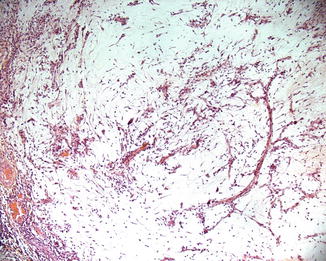
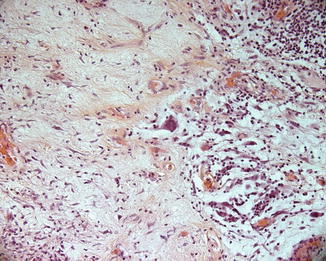
MFS is characterized by a proliferation of fusiform or stellate fibroblasts and delicate, thin-walled curvilinear blood vessels set within a prominent hyaluronic acid-rich myxoid stroma (Fig. 30.2). A prominent mixed inflammatory infiltrate may be seen (Fig. 30.3). It may exhibit a remarkably broad spectrum of histopathologic features ranging from low to high grade. Low-grade myxofibrosarcomas are hypocellular tumors, composed of relatively monomorphous cells. Careful examination invariably shows nuclear pleomorphism and hyperchromasia, but mitotic figures and other overtly malignant features may be inconspicuous (ca. 2 per 10 high-power field) in low-grade lesions (Fig. 30.4). It may contain cells resembling lipoblasts, sometimes referred to as pseudolipoblasts. This tumor cell subtype is filled with bubbly cytoplasmic vacuoles that compress and indent the nuclei in a manner similar to that seen in the lipoblasts that characterize liposarcomas. These cells are actually fibroblasts, and their vacuoles contain acidic glycosaminoglycans rather than the lipids of lipoblasts. Pseudolipoblasts may be a helpful diagnostic clue, but they are not invariably present, and several other tumors may produce morphologically similar cells.


Fig. 30.2
Myxofibrosarcoma presenting in the skin involves the reticular dermis, although the main bulk of the tumor is located in the subcutaneous tissue

Fig. 30.3
Myxofibrosarcoma is characterized by a proliferation of fusiform or stellate fibroblasts and delicate, thin-walled curvilinear blood vessels set within a prominent hyaluronic acid-rich myxoid stroma

Fig. 30.4
An infiltrate of atypical spindle cells within a prominent myxoid stroma and a pleomorphic multinucleated epithelioid cell component associated with a mixed inflammatory infiltrate may be seen
With increasing histological grade, myxofibrosarcomas show increasing cellularity, nuclear pleomorphism, and mitotic activity. Hemorrhage and necrosis can be present. Cells are usually spindle or ovoid but may also exhibit predominantly epithelioid cytologic features.
Immunohistochemically, tumoral cells are positive for vimentin and negative for desmin, pancytokeratin, CD31, CD34, CD68, and HMB45. S-100 can be positive in focal areas. In a minority of cases, some spindled or larger eosinophilic tumor cells express smooth muscle actin (SMA) and/or muscle-specific actin (MSA), suggestive of focal myofibroblastic differentiation. The myxoid matrix is composed of polysaccharide glycosaminoglycans such as hyaluronic acid, chondroitin sulfate, and keratan sulfate that stain positively with Alcian blue and not at all or very weakly with the periodic acid–Schiff (PAS) stain.
Stay updated, free articles. Join our Telegram channel

Full access? Get Clinical Tree


