Fig. 75.1
Skin metatstasis from a primary papillary thyroid carcinoma (courtesy of B.Cribier, Strasbourg, France)
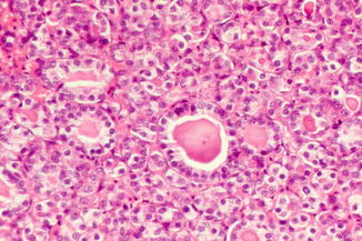
Fig. 75.2
The skin tumor is composed of tubules containing colloid-like material and lined by cells with nuclear grooves. (courtesy of B.Cribier, Strasbourg, France)
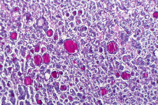
Fig. 75.3
The tumor is positive with PAS stain(courtesy of B.Cribier, Strasbourg, France)
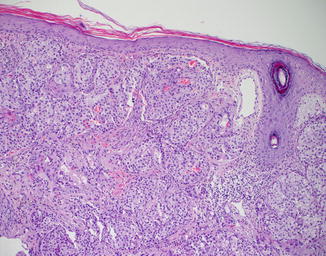
Fig. 75.4
Skin metastasis from a primary insular carcinoma, a follicular-related variant of thyroid carcinoma.Note solid cords of round or oval monotonous cells surrounded by fibrovascular septa (courtesy of W.Kempf, Zurich, Switzerland)
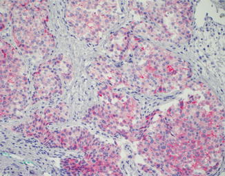
Fig. 75.5
Immunostaining reveals reactivity to thyroglobulins
Differential Diagnosis
Twenty-nine to 45 % of skin lesions are not suspected of being metastases due to an unusual clinical presentation. Common clinical diagnoses which are weighed are nonmelanoma skin cancer, cutaneous lymphoma, and cutaneous sarcomas.
Prognosis
Skin metastases from thyroid malignancies are manifestations of advanced disease, independently from the histological types. Age over 45 years and follicular pathology were significant predictors of a poorest outcome. In the setting of follicular carcinoma, patients with extracutaneous involvement at the time of cutaneous metastasis may have a worse prognosis than those with isolated cutaneous metastases. The average length of survival after cutaneous metastasis is 19 months.
Related posts:


Stay updated, free articles. Join our Telegram channel

Full access? Get Clinical Tree


