Fig. 51.1
Well-circumscribed, slowly growing lesion, 2 cm in diameter, with dark brown appearance, mimicking a melanocytic nevus
Pathology
“MDM” presents as an expansile nodule composed of relatively uniform, moderately atypical melanocytes. The cells may have an epithelioid or spindle cell appearance and exhibit only moderately enlarged nuclei with irregular chromatin distribution and an increased nuclear–cytoplasmic ratio. Mitotic figures are usually identified, but the mitotic index is quite low. There is no maturation of the cells or nests with progressive descent in the dermis.
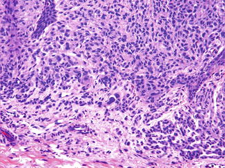
NM presents as a raised, dome-shaped, polypoid or papillomatous melanocytic proliferation, with or without a verrucous surface, mimicking a dermal nevus (Fig. 51.2). There is an apparent maturation in respect to the size of melanocytes and nests, with progressing descent in the dermis (Fig. 51.3). Frequently, the deep margin has an infiltrative growth pattern. The neoplasm is composed of a mildly pleomorphic population of melanocytes with hyperchromatic nuclei and prominent nucleoli (Fig. 51.4). There is an increased mitotic activity in the deeper aspects of the lesion. The intraepidermal melanocytes are arranged as both single cells and nests at the dermal–epidermal junction. Pagetoid spread of melanocytes may be present in a proportion of cases. Perineural infiltration may be occasionally encountered. Also, a peritumoral lymphocytic infiltrate is usually present.


Fig. 51.2
At scanning magnification, the lesion is large, polypoid, relatively symmetrical, and well circumscribed, reminiscent of a congenital melanocytic nevus. However, the high cellular density is the first clue that points to the real nature of the lesion

Fig. 51.3


Careful scrutiny of the lesion shows no real maturation and no stromal reaction
Stay updated, free articles. Join our Telegram channel

Full access? Get Clinical Tree


