Fig. 22.1
A slowly enlarging, flesh-colored, indurated plaque with ill-defined borders on the face
Pathology
At scanning magnification, MAC has a silhouette of a malignant neoplasm, being large, asymmetrical, and poorly circumscribed and usually extending deep into the dermis and subcutaneous fat and sometimes into the fascia, skeletal muscle, and bone (Figs. 22.3 and 22.4). The neoplasm has a characteristic appearance with cornifying cystic structures lined by squamoid cells in the superficial portion, solid aggregations of cells with pale or slightly dark eosinophilic cytoplasm in the middle part, and tubular structures lined by one or two layers of cuboidal cells that contain homogeneous eosinophilic material in the deeper aspects of the tumor (Fig. 22.2). These components are not always present and frequently may overlap. Some cases show a preponderance of solid structures with few if any cornifying cystic and tubular structures. Other cases show a predominance of tubular structures and may overlap with syringoid eccrine carcinoma. Tubules or cysts with tadpole-like morphology may occasionally be seen. There is no connection with the epidermis, but some examples show attachment to preexisting infundibula. Clear cell change, foci of decapitation secretion, sebaceous differentiation, and small whorls of compactly organized blue-gray corneocytes suggesting inner sheath differentiation may be seen.
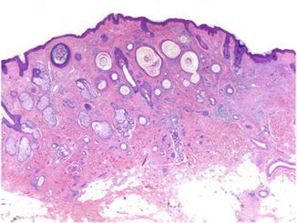
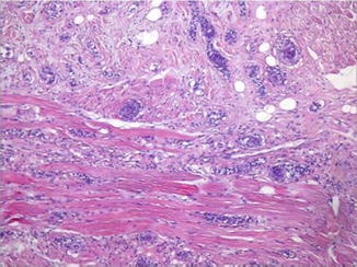
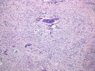
Fig. 22.2
The neoplasm has a characteristic appearance with cornifying cystic structures in the superficial part of it, and solid aggregations of epithelial cells admixed with tubular structures in its deeper aspects
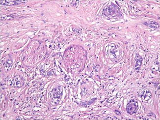
Fig. 22.3
The tumor usually extends into the subcutaneous fat and sometimes into the skeletal muscle. Tubules or cysts with tadpole-like morphology may occasionally be seen
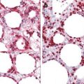
Fig. 22.4
MAC is notorious for its infiltrative and destructive growth. This neoplasm shows extension into the sphenoid sinus. The tumor is embedded into a desmoplastic stroma
The nuclei of the neoplastic cells are mainly small and monomorphous and usually lack nuclear atypia and mitotic figures. Frequently, the neoplasm manifests perineural involvement (Fig. 22.5). The epithelial component is disposed in a variably hyalinized stroma with thickened collagen bundles.