• The overall facial arc should have a slight convexity, with the projection greatest at the subnasale (anterior nasal spine [ANS]).
• Women typically prefer a more convex face, which is more feminine, rather than a flatter face, which is typical of increasing masculinity and preferred by men.
• Maxillary central incisor show at rest is 2 to 3 mm in males and 3 to 4 mm in females. During smiling, 1 to 2 mm of gingival show is considered ideal, whereas there should be no gingival show at repose. Increasing dental display gives a more youthful appearance to the face, and today, a slight gummy smile is considered more attractive. Maxillary gingival exposure decreases with lip lengthening, which occurs with aging.
• Incisor angulation and the maxillary dental arch are important for appropriate upper lip support and achieving an attractive smile arc. Because this can be controlled by the orthodontist with the individual dentition and the surgeon with a global movement, it is critical that both components are integrated.
• Computer-assisted planning allows the orthodontist and the surgeon to plan with precision and to be able to assess various options to optimize the aesthetic and functional components of the reconstruction. The simulation allows the surgeon to design osteotomies that are specifically tailored to the patient and to assess the outcomes of the various options and intraoperative points of difficulty.
• When conventional orthognathic surgery cannot provide the desired aesthetic projection and symmetry, alloplastic implants can improve the desired surface contours as a secondary staged procedure.
Introduction
Among the essential procedures in the surgical armamentarium of an aesthetic maxillofacial surgeon is sectioning and repositioning of the midfacial skeleton. The versatility of the LeFort I surgery and its variations allows the surgeon to correct a broad spectrum of midfacial skeletal abnormalities. The maxilla forms the most central portion of the facial skeleton and contributes significantly to the overall aesthetic appearance of the face. Deficiency or excess of the maxilla in the vertical or sagittal dimension can profoundly affect midface concavity/convexity, the occlusal plane, dental and gingival show, the smile arc and lip support, nasal dorsal prominence and nasal tip position. The spatial position of the maxilla relative to the cranial base and its counterpart, the mandible, can therefore have a significant impact on facial aesthetics and dental occlusion. This chapter focuses on the surgical techniques for repositioning the maxilla to restore and enhance aesthetic and dental harmony.
Maxillary skeletal deformities
Abnormalities of the maxilla in the vertical dimension may include deficient maxillary height. This results in decreased show of the anterior teeth, counterclockwise rotation of the mandible, and associated soft tissue changes that result in a prematurely aged look to the face. Vertical maxillary excess can produce excess show of the anterior teeth and an undesirable, gummy smile. The excess height of the maxilla also results in clockwise rotation of the mandible and associated soft tissue changes, such as lip incompetence, mentalis strain, and a retruded chin.
Abnormalities of the maxilla in the anteroposterior, sagittal dimension may result in negative or excess overjet if there is deficient or excess sagittal projection of the maxilla, respectively. Maxillary deficiency in this dimension also results in an acute nasolabial angle, nasal dorsal prominence with a depressed tip and narrow alar base, decreased dental show with smiling, concavity of the midface, perceived relative prognathism, and a short retrusive upper lip with thin vermilion.
Patient assessment
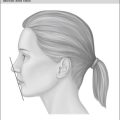
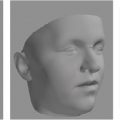
When asked about their appearance, patients frequently describe their concerns in terms of the mandible. It is not uncommon for a patient with a class III facial skeletal pattern to request setting back the mandible ( Fig. 20.1 ). The clinical clues that the surgeon needs to point out to the patient with maxillary deficiency include the acute nasolabial angle, nasal dorsal prominence with depressed nasal tip, lack of dental display, shallow piriform region, and midfacial concavity. When reviewing photographs with the patient, it is the three-quarter oblique view that best shows the midfacial concavity, which can extend to involve the zygoma ( Fig. 20.2 ). Even when the primary deformity is in the mandible as in “true” mandibular excess and prognathism, maxillary advancement should be considered to improve the overall aesthetic appearance because it expands the facial skeleton to accommodate the soft tissue envelope, thus countering prematurely aged appearance associated with skeletal contraction. Mandibular surgery combined with maxillary surgery often improves postoperative stability and improves the aesthetic outcome. Thoughtful clinical examination without skeletal radiography will, in most circumstances, appropriately define the maxillary and mandibular components to optimize the aesthetic outcome. However, dental models and radiographs become essential in detailing the surgical–orthodontic plan to optimize the skeletal aesthetic outcome and occlusal function.
Evaluation includes the facial soft tissue, the skeletal bases, dental occlusion, and the position of the dental arches relative to that of the lips. The respective heights of the upper, middle, and lower face should be noted. Facial convexity or concavity should be assessed. The frontonasal angle, nasal length, columella height, and nasolabial angle should be determined. Smile analysis should be performed to determine the lip height at rest, the smile arc and lip dynamics, lip competence, and the presence of mentalis strain. The incisal edges of the maxillary dentition should ideally follow the lower lip (smile arc). The molar and canine relationships of the maxilla and the mandible should be assessed relative to the goals of optimizing the skeletal appearance. Thus the degree of the overjet, overbite, incisor inclinations, and occlusal plane angulation should be carefully assessed by both the surgeon and the treating orthodontist to optimize the facial and occlusal aesthetic appearances.
Objective records
Today, the more conventional orthopanorex, the frontal and lateral cephalograms, are being replaced by cone beam computed tomography (CBCT) scans, which provide a more complete skeletal anatomy in three dimensions, allowing the surgeon and the orthodontist to fully assess the facial skeleton in multiple dimensions avoiding the inherent errors of registering a series of two-dimensional radiographs. Similarly, there is an increasing trend toward three-dimensional occlusal surface scanning to avoid the inconvenience of obtaining dental impressions and plaster/stone fabrication. Finally, there are increasingly more affordable options of three-dimensional surface imaging to replace a series of conventional two-dimensional photographs of the patient.
There is, today, a multitude of software products allowing the surgeon and the orthodontist to create a virtual three-dimensional “head” as a composite of the surface images, the underlying skeleton, and the dentition. This digital model allows for integrated surgical and orthodontic treatment planning, with a precision that could not be achieved with conventional analog planning.
Treatment goals and orthodontic and surgical planning
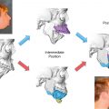
From an aesthetic point of view, the surgeon needs to determine the final position of the midfacial skeleton relative to the fronto-orbital (skeletal base) region and the final position of the mandible (body and chin). The dentition needs to be then repositioned by the orthodontist working with the surgeon to achieve the desired skeletal aesthetic position. In circumstances where there is insufficient dental discrepancy to achieve the desired maxillary movement, premolars may need to be extracted to create an appropriate overjet with a class I canine relationship regardless of the molar Angle classification. Additionally, the final position of the dental arches and the incisor inclinations need to be determined to optimize the dental display in repose and during smiling and the smile arc. Because of the multitude of variables that need to be controlled, the surgeon and the orthodontist must work together to achieve the patient’s desired goals by using digital tools that provide the precision needed. Effective preoperative planning will optimize postoperative outcomes. Virtual surgical planning with computer-aided design/computer-aided manufacturing (CAD-CAM) can help depict the desired skeletal and dental movements and to generate preoperative orthodontic arch wires and intraoperative surgical guides ( Fig. 20.3 ). This is detailed in Chapter 17 on virtual surgical planning.