Fig. 37.1
(a) Photograph of the maxilla allograft just after anastomoses. Please note that one side artery (marked with “a”: end-to-end, femoral to common carotid) an done side vein anastomosias (marked with “v”: end-to-end, external jugular to femoral) The After anastomosis artery on venous anastomosis side and vein on artery anastomosis side was ligated. (b) One hundred five days after transplantation intact vascular pedicles marked with a and v can be seen. The venous cannula was inserted to the posterior opening of the nasal passage (coana)
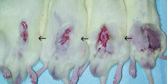
Fig. 37.2
Sequential phots of the grafts postoperatively. Starting from right side of the photo: 2nd day after transplant, 2 weeks after transplant in which graft was exteriorized, 1 month after transplant and 105th day after transplant (left end)
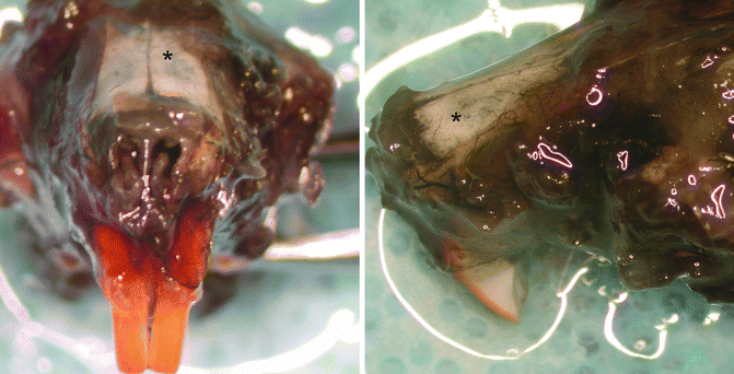
Fig. 37.3
Photos of the maxilla graft after indian ink angiography via carotid pedicle and glycerol clearence, from anterior and left side. Asterisk shows the perfusion of the bone through periosteal supply
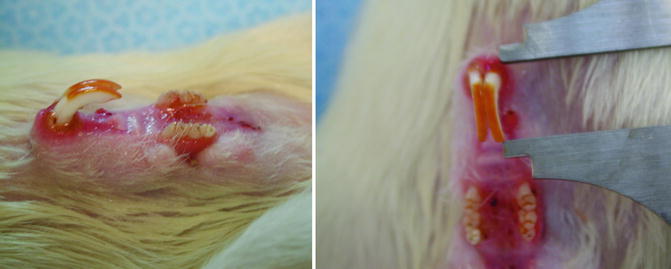
Fig. 37.4
Photos of the maxilla graft after105 days postoperatively. From lateral and anterior views. The incisive teeth was measured,1.2 cm long
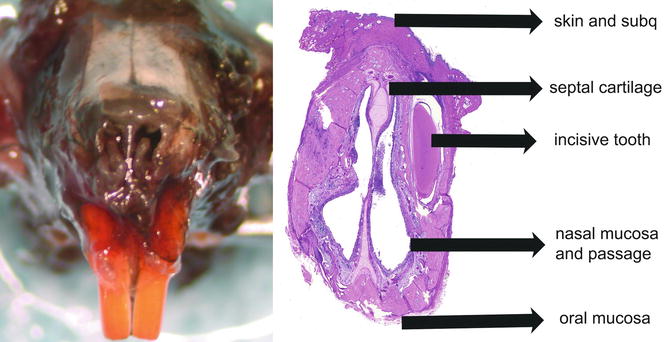
Fig. 37.5
 Microcirculation Model for Invasive Animal Monitoring
 Composite Osseomusculocutaneous Thymus Allotransplantation Model
 In Vivo Chimera Model: Creation of Primary and Secondary Chimera
 Peripheral Nerve Surgery Models Crush Injury and Epineural Patch
 Heterotopic Vascularized Ovarian Autotransplantation Model in the Sheep
 Defect Repairs After Resections of Laryngeal Cancer, Hypopharyngeal Cancer, and Cervical Esophageal Cancer


Related posts:
Microcirculation Model for Invasive Animal Monitoring
Composite Osseomusculocutaneous Thymus Allotransplantation Model
In Vivo Chimera Model: Creation of Primary and Secondary Chimera
Peripheral Nerve Surgery Models Crush Injury and Epineural Patch
Heterotopic Vascularized Ovarian Autotransplantation Model in the Sheep
Defect Repairs After Resections of Laryngeal Cancer, Hypopharyngeal Cancer, and Cervical Esophageal Cancer
Stay updated, free articles. Join our Telegram channel
Full access? Get Clinical Tree
