The task of managing an open wound complicated by exposed bony structures underneath is difficult, if not challenging. We have instituted a method of managing the problems in stages using an artificial dermis and skin grafting technique in 17 wounds in 15 individuals from Sept. 2006 to Feb. 2009. While all wounds were noted to assume aberrant healing processes, the majority of involved bony structures were devoid of periosteal covering compounded by various degrees of infection. Of 15 incidents, mechanical trauma was responsible for 10, chemical burns for two and electrical burns for two patients. A chronic non-healing ulcer with exposed bone formed in an old burn scar accounted for the remaining one. The regimen of surgical management consisted of initial debridement, the coverage of the resultant wound with an artificial dermis and a partial-thickness skin grafted over this dermis-like structure grown with granulation tissues. Complete wound healing was attained in 15 out of 17 with outstanding cosmetic and minimal donor-site morbidity. Despite initial failure encountered in two, the morbidities noted were low. It is especially useful in large defects that usually require flaps for coverage.
Problems associated with a non-healing wound are frequently noted in individuals with mechanical trauma and burns, pressure ulcers and diabetic illness. The task of managing such a wound, especially in instances with bony exposure, is clinically challenging, if not difficult.
Most treatment approaches involve conservative treatment and surgery. Currently, surgical treatment often uses local or distal skin flaps, muscle flaps or myocutaneous flaps to repair defects.
The most simplistic approach in managing a wound that could not be closed primarily is to use a piece of skin graft for coverage. Although a skin flap mobilised from the area adjacent or from a distant site has been advocated to manage a wound with vital structures exposed, that is, bone, vessels and nerves, the techniques in practice are often plagued with problems such as paucity of flap donor sites. The magnitude of morbidities associated with these procedures further precludes their use.
Artificial dermis, with its silicone membrane, collagen–sponge bilayer structure, was the first tissue-engineered skin replacement that could be successfully used in clinical situations. Artificial dermis is mainly used to repair skin and soft tissue defects; it has also been used to successfully repair wounds with partial tendon exposure even with exposed bone wound.
A regimen of wound management that includes the initial coverage of a newly freshened wound with an artificial dermis and grafting of the resultant wound with a piece of autologous partial-thickness skin graft was tried at our hospital in 17 wounds in 15 patients. The experience gained from managing this group of patients formed the basis of this article.
Clinical materials and methods
Patient Materials
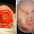
A total of 15 inpatients were treated during 2.5 years. There were 11 men and four women. While the youngest was 6 years, the oldest was 72 years, with a mean age of 38.14 ± 17.29 years. As noted in the Table 1 , all wounds were located in the lower extremities and all had the underlying bony structures exposed Fig. 1 . Mechanical trauma noted in 10 patients was the most common cause of the wounds while chemical burns in two and electrical burns in one were the causes. Breakdown of an old burn scar was the cause in the other patient.
| Patient | Age (y) | Gender | History Summary | Bone-exposed Part and Size | Wound Infection | Surgical Bed | Interval Between Surgeries (days) | Outcome | Follow-up Period (months) |
|---|---|---|---|---|---|---|---|---|---|
| 1 | 72 | Female | After steel plate internal fixation of right lateral malleolus fracture, the steel plate was exposed for 6 weeks. Patient had a >10-y history of diabetes. | Right lateral malleolus, 2 cm × 5 cm, exposed wound | Yes | Bone | 20 | Healed | 18 |
| 2 | 43 | Male | Left distal tibial open fractures with exposed ankle joint for 9 weeks. | Left distal tibial, 2 cm ×2.5 cm, exposed wound | Yes | Bone and exposed joint | Infected | Failed | 18 |
| 3 | 39 | Male | Wheel crush injury to the right leg. Severe skin abrasion and muscle laceration; tibia partially exposed for 2 weeks. | Right proximal tibia, 3 cm × 5 cm, exposed wound | Yes | Bone | 16 | Healed | 12 |
| 4 | 34 | Female | Right foot dorsum burn scar ulceration with exposed bone, 15-y history of repeated ulcerations. | Right foot dorsum metatarsals, 2 cm × 2.5 cm, exposed wound | No | Bone | 16 | Healed | 11 |
| 5 | 21 | Male | Widespread skin and soft tissue necrosis after anhydrous ethanol injection for right leg lymphocele, 6-wk history. | Right proximal tibia, 3 cm × 18 cm, exposed wound | Yes | Bone | 28 (First implantation) 15 (Second implantation) | Healed | 10 |
| 6 | 33 | Male | Right distal tibial and fibular open fractures, 38 days after external frame fixation. | Right tibia 3 cm × 5 cm, exposed wound, exposure of fracture ends | Yes | Bone | 28 | Healed | 8 |
| 7 | 6 | Male | Wheel crush injury to the right leg. Femur and tibia fracture, severe skin abrasion and necrosis; tibia partially exposed for 19 days. | Right tibia 1.5 cm × 3 cm, exposed wound | Yes | Bone and periosteum | 15 | Healed | 5 |
| 8 | 30 | Female | Wheel crush injury to the right lower leg and foot. Severe skin abrasion and muscle laceration; tibia, 5th dorsum metatarsal and calcaneus partially exposed for 26 days. | Right tibia 1 cm × 3 cm | Yes | Bone and periosteum | 14 | Healed | 8 |
| Right 5th dorsum metatarsal 0.8 cm × 1 cm | 14 | Healed | |||||||
| Right calcaneus 3 cm × 3.5 cm, exposed wound | Infected | Failed | |||||||
| 9 | 28 | Male | High voltage electronic burn injury to left foot, calcaneus and achilles tendon exposed for 6 weeks. | Left calcaneus 2 cm × 2.5 cm, exposed wound | Yes | Bone and periosteum | 24 | Healed | 4 |
| 10 | 39 | Male | Chemical burn to lower legs and feet; dorsum metatarsals and achilles tendon partially exposed for 9 weeks. | Left first dorsum metatarsal, 1 cm × 2 cm, exposed wound | Yes | Bone | 21 | Healed | 2 |
| 11 | 36 | Male | Crush injury to lower extremity; right thigh amputation; Severe skin avulsion to left lower leg; tibia partially exposed for 19 days. | Left tibia anterior part 3.5 cm × 22 cm, exposed wound | Yes | Bone | 21 | Healed | 6 |
| 12 | 72 | Female | Chronic ulcer to right knee with tibial tubercle exposed for 2 years. | Right tibial tubercle 1.5 cm × 1.5 cm exposed wound | No | Bone and periosteum | 18 | Healed | 2 |
| 13 | 38 | Male | Traffic accident crush to the right leg, excision of external malleolus after infection, chronic ulcer on external malleolus for 3 years, deep vein thrombosis | Right distal fibula 2.5 cm × 1.5 cm exposed wound | Yes | Bone and periosteum | 20 | Healed | 1 |
| 14 | 43 | Male | Chronic ulcer on left medial malleolus for 17 years. | Left medial malleolus 1.5 cm × 1.5 cm exposed wound | Yes | Bone and periosteum | 14 | Healed | 1 |
| 15 | 29 | Male | High voltage electronic burn injury to right leg and foot for two days. | Left tibia anterior part 3.8 cm × 12 cm, exposed wound | No | Bone and periosteum | 21 (First implantation) 21 (Second implantation) | Healed | 1 |
Related posts:
 Stiffening of Human Skin Fibroblasts with Age
 Physiology of Skin Aging
Tissue Engineering of Skin
Physiology of Skin Aging
Tissue Engineering of Skin
 Dermal Substitutes Do Well on Dura: Comparison of Split Skin Grafting +/− Artificial Dermis for Reconstruction of Full-thickness Calvarial Defects
Management of Split Skin Graft Donor Sites–Results of a National Survey
Skin: Histology and Physiology of Wound Healing
Dermal Substitutes Do Well on Dura: Comparison of Split Skin Grafting +/− Artificial Dermis for Reconstruction of Full-thickness Calvarial Defects
Management of Split Skin Graft Donor Sites–Results of a National Survey
Skin: Histology and Physiology of Wound Healing
Stay updated, free articles. Join our Telegram channel

Full access? Get Clinical Tree


