Fig. 58.1
In this case of MPNST, the skin involvement is secondary to local invasion from a subcutaneous tumor
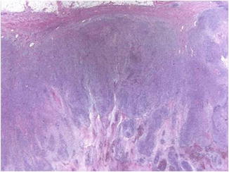
Fig. 58.2
The tumor is large, asymmetrical, and unencapsulated
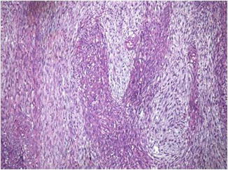
Fig. 58.3
Irregular interlacing bundles of spindle cells display a “marbled” pattern with hypercellular fascicles of spindle cells alternating with hypocellular myxoid areas
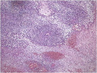
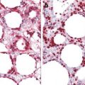
Fig. 58.4
Focal areas of perivascular cuffing by tumor cells is a typical feature
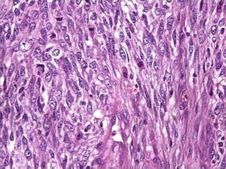
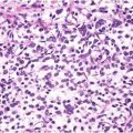
Fig. 58.5
The spindle cells have hyperchromatic, pleomorphic wavy nuclei, with high mitotic activity
MPNST with spindle cell morphology is characterized by a focal and week positivity reaction for S-100. In contrast, the epithelioid variant stains stronger and more uniform with S-100 and is also positive for podoplanin (D2-40). MPNST shows a positive reaction for vimentin, myelin basic protein (MBP), CD56, and CD57 (Leu7) (Fig. 58.6a–c) and a negative reaction for HMB45, melan-A, tyrosinase, microphthalmia transcription factor, smooth muscle actin, and cytokeratin. Proliferative index with Ki67 is high (Fig. 58.6d).
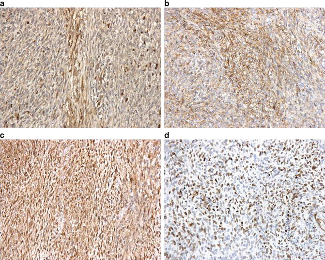
Fig. 58.6


The neoplastic cells are (a) focally positive for S-100, (b) diffusely positive for CD56, and (c) diffusely positive for PGP 9.5 and (d) show a high index of proliferation with Ki67
Related posts:


Stay updated, free articles. Join our Telegram channel
Full access? Get Clinical Tree
