Fig. 18.1
Malignant mixed tumor displays dermal and/or subcutaneous nodules composed of epithelial cells arranged in cords and nests within a chondromyxoid stroma
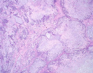
Fig. 18.2
The tumor has a multinodular architecture and prominent chondromyxoid stroma. Ductal differentiation is present in the lower left of the image
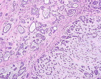
Fig. 18.3
The epithelial cells are arranged into nests with ductal differentiation or into cords and strands set within a chondromyxoid stroma
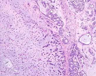
Fig. 18.4
The stroma may bear a remarkable similarity to cartilage
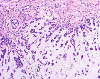
Fig. 18.5
The epithelial cells are arranged into nests with ductal differentiation or into cords and strands set within a chondromyxoid stroma
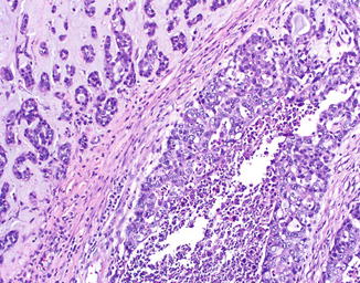
Fig. 18.6
Marked nuclear atypia and focal tumor necrosis may be present
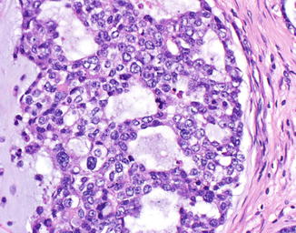
Fig. 18.7
Nuclear pleomorphism and atypical mitoses may be seen, although some cases of malignant mixed tumor are deceptively banal
Differential Diagnosis
The main histological differential diagnosis includes benign mixed tumor for banal-appearing malignant mixed tumors and other forms of poorly differentiated carcinoma for markedly atypical-appearing malignant mixed tumors. Other entities in the differential diagnosis include mucinous eccrine carcinoma, sarcomatoid carcinoma (carcinosarcoma) with chondrosarcomatous areas, matrix producing melanoma, metastatic chondrosarcoma, chondroma of soft parts, extraskeletal myxoid chondrosarcoma, ossifying fibromyxoid tumor of soft parts, and extra-axial soft tissue chordoma. Myoepithelioma and myoepithelial carcinoma have similar features to both benign and malignant mixed tumors; myoepithelial neoplasms and mixed tumors may exist on a spectrum.
Stay updated, free articles. Join our Telegram channel

Full access? Get Clinical Tree


