Fig. 21.1
A rapidly growing, firm in consistency, reddish-blue nodule on the forearm
Pathology
Histopathological diagnosis of MEC requires the detection throughout the tumor, of at least one focus, variably represented, of benign eccrine spiradenoma, with the typical dual cell population, arranged in nests and trabeculae (Figs. 21.2 and 21.3). The malignant features are not specific and include solid aggregates of tumor cells with mild to moderate nuclear atypia, loss of two cell populations, an infiltrative pattern of growth, atypical mitotic figures, necrosis, lymphovascular, and perineural invasion (Figs. 21.4 and 21.5). Other features are invasion of surrounding connective tissue, loss of basement membrane, squamous differentiation, and clear cell, oncocyte-like, and sarcomatoid changes. “Sarcomatous” or “squamous” changes seem significantly to be correlated with a poorer prognosis. Two distinct morphological patterns of MEC have been described, the most common form consisting of an obviously malignant pleomorphic tumor (high-grade carcinoma) and a second low-grade form that closely mimics benign spiradenoma. Some malignant tumors showing features of both spiradenoma and cylindroma are named “spiradenocylindrocarcinoma.” These features support the evidence that both tumors are derived from a common pluripotential basal cell line.


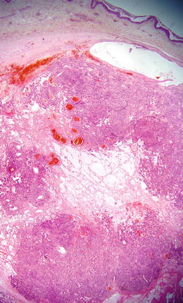
Fig. 21.2
A basaloid nodular proliferation without epidermal connection

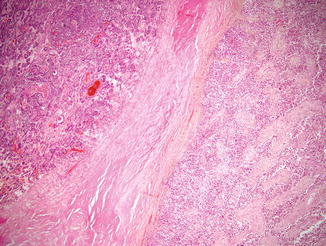
Fig. 21.3
An area with carcinoma (right) adjacent to a benign eccrine spiradenoma (left)

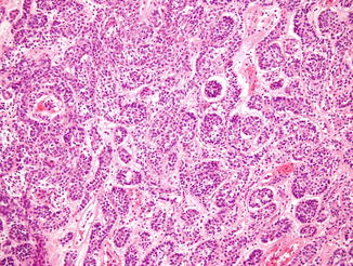
Fig. 21.4
The tumor is composed on nests and cords with loss of the dual population

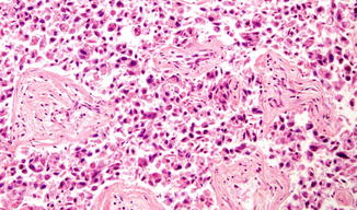
Fig. 21.5


Malignant features with aggregates of tumor cells with mild to moderate nuclear atypia, necrosis, lymphovascular, and perineural invasion
Stay updated, free articles. Join our Telegram channel

Full access? Get Clinical Tree


