| Reconstruction after excision of eyelid neoplasm |
| Focal trichiasis refractory to epilation or lash follicle destruction |
| Need for lower eyelid tightening |
| Repair of traumatic eyelid laceration |
| Correction of irregular eyelid margin contour (congenital or secondary to prior surgery such as eyelid margin destruction after cryotherapy) |
| History of trauma, prior surgery, cancer |
| Involvement of punctum/proximal lacrimal drainage system, particularly for neoplasms and trauma |
| Biopsy of any suspicious lesions |
| Anterior/posterior lamellar deficiencies |
| Associated eyelid malpositions – ectropion/entropion/eyelid retraction/lagophthalmos |
| Degree of lower eyelid laxity |
Introduction
Wedge resection of the eyelid can be utilized for removal of diseased segments of the eyelid from neoplasm and trichiasis and even for tightening of eyelid laxity. Most commonly, wedge resection with reconstruction of eyelid is used for reconstruction after removal of cutaneous malignancies.
Eyelid lesions can range from benign cysts and inflammatory lesions (hordeolums/chalazions) to malignancies. Although clinical examination can be extremely helpful in diagnosing typical eyelid lesions (cysts, nevi, papillomas), other more atypical lesions are often hard to differentiate by clinical exam alone. Furthermore, certain conditions such as sebaceous cell carcinoma can masquerade as a chalazion or chronic blepharitis.
Approximately 10% of all skin malignancies present on the eyelid. Of these, the highest incidence is basal cell carcinoma, followed by squamous cell carcinoma, sebaceous cell carcinoma, and malignant melanoma. Biopsy with pathological analysis is the only definitive way to determine the etiology of an unknown eyelid lesion.
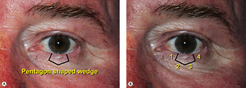
Generally, a lesion involving one-third of the eyelid margin or less can be approached using wedge resection and direct closure/reconstruction of the eyelid. Defects greater than 50% may require a semicircular or pedicle-based flap ( Chapter 38 , Chapter 39 , Chapter 40 , Chapter 41 , Chapter 42 , Chapter 43 ). A complete history is necessary, including the chronicity of a lesion, associated symptoms, discharge, pain, bleeding, and family history of skin malignancies. In trauma and lacerations, it is important to determine the mechanism of injury and if the tetanus vaccine status is current. It is also prudent to determine if the patient is on anticoagulants or has a clotting disorder.
A complete ocular examination is also necessary. One should examine the eyelid margins and lesion for size, depth, extent, involvement of anterior/posterior lamellae, bulbar/palpebral conjunctival, punctal, canalicular and lacrimal drainage system. Furthermore, one should examine for madarosis, trichiasis, vascularization, irregularities, pigmentation, ulceration, entropion, ectropion, and eyelid laxity. Lymph nodes (including pre-auricular, submandibular, and cervical nodes) should be palpated for any evidence of metastases. Photographic documentation of the eyelid lesion before biopsy, intraoperatively and postoperatively are highly recommended.
A meticulous, layered closure with restoration of normal anatomy is essential to maximize form and function after wedge excision and reconstruction. The eyelid margin is repaired with silk sutures which provide strength and induce enough inflammation to ensure adequate healing while minimizing irritation of the ocular surface.
Surgical Technique
Related posts:


Stay updated, free articles. Join our Telegram channel

Full access? Get Clinical Tree


