Lower eyelid reconstruction with Mustardé flap


Table 41.1

Indications for surgery


Lower eyelid defects up to 100% total eyelid
Large eyelid–cheek junction skin defects
Large cheek skin defects


Table 41.2

Preoperative evaluation


History of malignant neoplasm of skin
History of smoking/tobacco use
History of prior facelift or periocular surgery
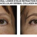
Presence of lower eyelid malpositions (ectropion, entropion, retraction, lagophthalmos)
Size and depth of soft tissue defect


Introduction


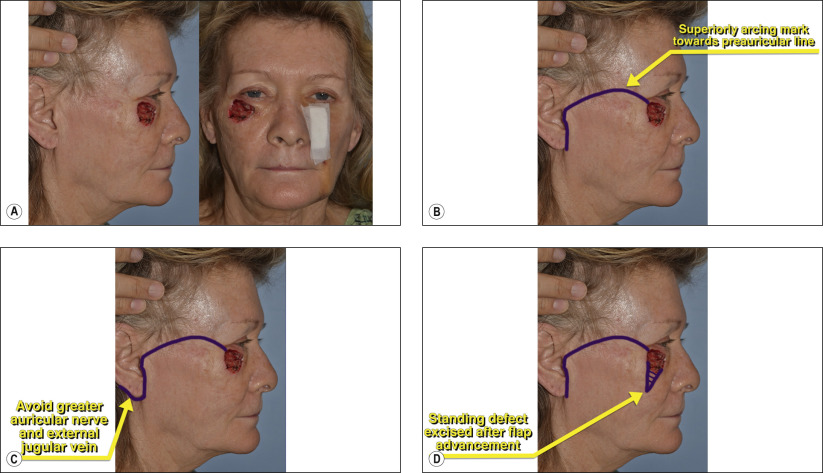
Mustardé’s cheek rotational flap is a versatile procedure to close large defects of the lower eyelid, eyelid–cheek junction and cheek commonly seen after the removal of cutaneous malignancies. In addition to supplying ample anterior lamella for functional reconstructions, Mustardé’s flap can result in a marked cosmetic improvement over a full or split thickness skin graft.


When performing a reconstruction of full thickness lower eyelid defects, the posterior lamella can be provided by an upper eyelid tarsoconjunctival flap ( Chapter 40 ) or a free tarsal graft from the contralateral eyelid ( Chapter 43 ) with the anterior lamella supplied by the Mustardé flap.


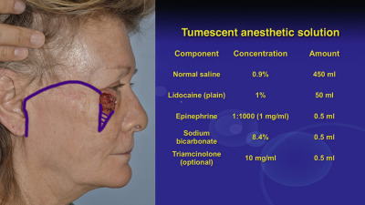
The Mustardé flap is performed analogously to the MACS facelift covered in Chapter 24 . A superiorly arcing mark is made towards the preauricular line and then a subcutaneous flap is developed. Extensive undermining is performed and the flap is rotated medially to close defects. At the base of the excision, a Burow’s triangle is excised to remove a redundant dog ear of tissue.


Postoperatively, patients are instructed to adhere to the same restrictions as facelift patients. Strict avoidance of tobacco is required, as this will cause vasoconstriction and potential necrosis of the flap. Patients are given postoperative antibiotic prophylaxis and, in select patients, a drain can be placed which is removed at 24 hours postoperatively.


Surgical Technique


Figures 41.1A–D


Skin marking

Figure 41.1A shows an eyelid–cheek junction defect over 40 mm in diameter after excision of a melanoma with permanent tissue margins. Reconstructive options include full or split thickness skin grafting or an adjacent tissue transfer flap. The disadvantages of the skin graft include color/textural differences, depression at the site of the graft, donor site morbidity and potential for graft loss. This defect is ideally suited to reconstruction with a Mustardé flap. Skin incision placement is demonstrated with a superiorly arcing mark from the upper lateral skin defect to the preauricular line ( Figure 41.1B ). If larger defects are present, the incision can extend posteriorly behind the ear lobe for a more comfortable rotation ( Figure 41.1C ). When dissecting posterior to the ear lobe, care is taken to avoid the greater auricular nerve and external jugular vein, which run superficial to the sternocleidomastoid muscle. Once the rotational flap is advanced medially, a standing defect will typically be present inferiorly ( Figure 41.1D ). This can be excised after the flap is advanced to minimize unnecessary tissue removal.


Figure 41.2


Tumescent anesthesia

The use of tumescent anesthesia provides excellent anesthesia coupled with a vasoconstricted surgical field. A tumescent solution is prepared by discarding 50 ml of saline from a 500 ml normal saline IV bag and adding the components shown in Figure 41.2 . This mixture will yield 500 ml of a dilute anesthetic solution that is liberally infiltrated into the entire flap at least 10 minutes prior to surgery. The tumescent solution can be given using a 10 ml syringe and a bent 22-gauge spinal needle. The Mustardé rotational flap can be performed using monitored anesthesia care or general anesthesia based on patient and surgeon preference.

Only gold members can continue reading. Log In or Register to continue

Stay updated, free articles. Join our Telegram channel

May 16, 2019 | Posted by in Reconstructive surgery | Comments Off on Lower eyelid reconstruction with Mustardé flap

Full access? Get Clinical Tree

Get Clinical Tree app for offline access