Key Words
rotation flap, transposition flap, advancement flap, bilobed flap, Z-plasty, Limberg flap
Synopsis
Local skin flaps are time-honored methods of soft tissue reconstruction and frequently represent the ideal mode of reconstruction because they permit defect coverage with skin of similar color, thickness, and texture. Successful reconstruction using local skin flaps, however, requires a profound understanding of cutaneous vascular anatomy, skin biomechanics, and tissue geometry. Pre-operative errors in flap choice and design can substantially mitigate the likelihood for a successful reconstruction. Meticulous pre-operative analysis of a skin defect, precise planning, and atraumatic surgical technique are prerequisites for a favorable outcome. This chapter discusses key concepts of local skin flap design, highlights surgical principles, and provides clinical examples to permit imminent translation into clinical practice.
Clinical Issues
The “reconstructive ladder,” which was introduced in 1982, continues to serve as the conceptual framework that allows plastic surgeons to decide on the appropriate reconstructive modality for any given defect. Whereas healing by secondary intention and wound coverage using skin grafts rely on an appropriately vascularized wound bed, lack thereof mandates the use of flaps for successful reconstruction. The differentiating factor between a graft and a flap is that the latter maintains its perfusion during transfer, thus making it independent from the vascularity at the recipient site. Typical indications for flap coverage include exposed tendons, bones, or joints. Local skin flaps may also be used to reconstruct skin defects in highly visible areas such as the face after skin cancer excision.
Flaps can be categorized in a variety of ways; for example, based on their blood supply, method of movement, proximity to the defect, configuration, and composition ( Table 2.5.1 ). The focus of this chapter is on local skin flaps, defined as flaps that are located in the vicinity of an existing defect. The blood supply to these flaps can be either random or axial, depending on their design and location. The most reliable flaps that can be used anywhere in the world are highlighted.
| Characteristic | Examples |
|---|---|
| Blood supply | Random pattern flap Axial pattern flap |
| Method of movement | Advancement flap Rotation flap Transposition flap |
| Proximity to the defect | Local flap Distant flap Free flap (microvascular tissue transfer) |
| Configuration | Rhomboid Bilobed |
| Composition | Cutaneous flap Fasciocutaneous flap Musculocutaneous flap |
Random pattern flaps with fixed length-to-width (such as 2 : 1 or 3 : 1) ratios were historically the most common type of flap transfer. The reason for these rigid ratios was an incomplete understanding of flap perfusion. In random pattern flaps, blood vessels contained within the flap were considered to be randomly oriented. In other words, random pattern flaps were raised without regard to the underlying vascular anatomy. The concept of fixed length-to-width ratios, however, was disproved as knowledge of vascular anatomy increased. In contrast to random pattern flaps, axial pattern flaps are raised based on an anatomically named vessel that courses along the longitudinal axis of the flap; examples include the forehead flap (supratrochlear artery) or the groin flap (superficial circumflex iliac artery). This allows flap harvest without the restrictions of fixed length-to-width ratios.
While advances in plastic and reconstructive surgery have resulted in an increase in the number of complex reconstructions being performed, including a rise in the number of microsurgical reconstructions, use of local skin flaps remains a major reconstructive modality with minimal donor site morbidity. Local skin flaps are raised from the tissue in the immediate vicinity of the defect and provide an ideal reconstructive solution because they are similar in color and texture to the skin at the site of the defect. They are transferred as either advancement, rotation, or transposition flaps. An in-depth discussion of all the local skin flaps available is beyond the scope of this chapter. As such, treatment principles and distinct clinical problems will be highlighted to discuss the utility of certain local skin flaps.
Clinical problems commonly treated with local skin flaps can be grouped into distinct categories, including:
- •
Tumor
- •
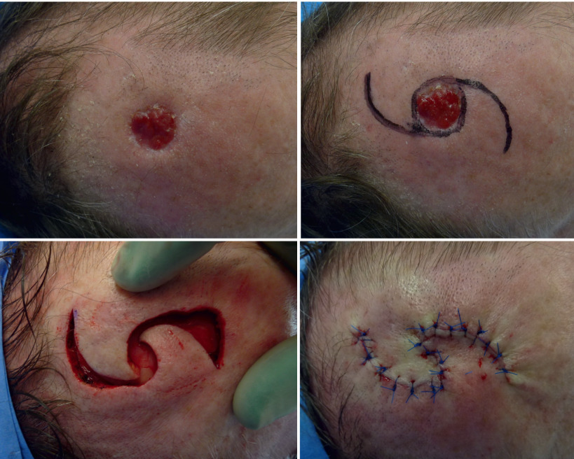
For example, rotation or transposition flaps for reconstruction after excision of skin cancer of the head and neck region ( Fig. 2.5.1 )
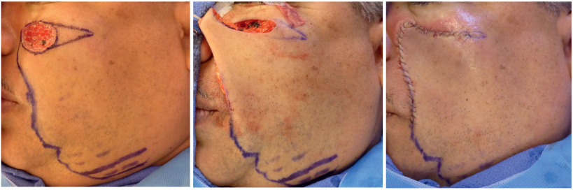
Fig. 2.5.1
Use of a local skin flap to reconstruct a facial defect after excision of a basal cell carcinoma.
- •
- •
Trauma
- •
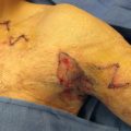
For example, V-Y advancement flap after distal finger injury or Moberg advancement flap after distal thumb injury ( Fig. 2.5.2 )
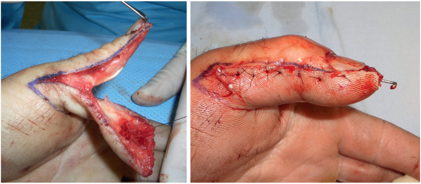
Fig. 2.5.2
Moberg advancement flap after distal thumb injury.
- •
- •
Congenital
- •
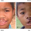
For example, four-flap Z-plasty for release of a first web space contracture ( Fig. 2.5.3 )
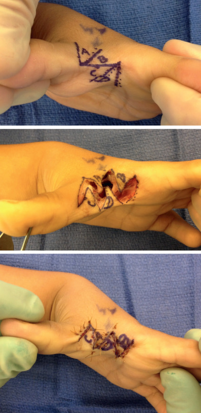
Fig. 2.5.3
Four-flap Z-plasty for contracture release of first web space.
- •
Management
Certain principles need to be adhered to before moving forward with reconstruction. In oncological cases, confirming complete tumor excision is mandatory. In traumatic wounds, adequate debridement before defect coverage is critical. In cases of contracture release, complete release of the scar is crucial to being able to adequately assess the true extent of the soft tissue defect and to adequately correct the deformity. Once these issues have been adequately addressed, one may proceed with the reconstruction.
Wound assessment should include determining the size of the defect as well as evaluating the area of skin and soft tissue redundancy in the vicinity of the defect. When designing a flap, it is important to plan a flap of sufficient dimension. Planning the local skin flap somewhat larger than the defect is prudent to avoid tension upon flap transfer and closure. Ideally, the pivot point of the flap should be determined pre-operatively.
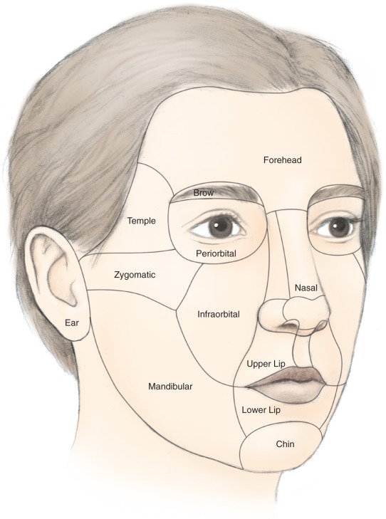
In flap design, understanding the surrounding anatomical structures and features is key to maintaining form and function. Commonly, numerous flap options exist for any given defect. Thus it is important to analyze location and direction of local skin laxity as well as tissue characteristics such as elasticity, thickness, and sensitivity to optimize the reconstructive outcome. For facial defects, keeping the flap within only one facial esthetic unit and designing incisions along relaxed skin-tension lines or at the edge of an esthetic unit will yield the best cosmetic outcomes ( Fig. 2.5.4 ).
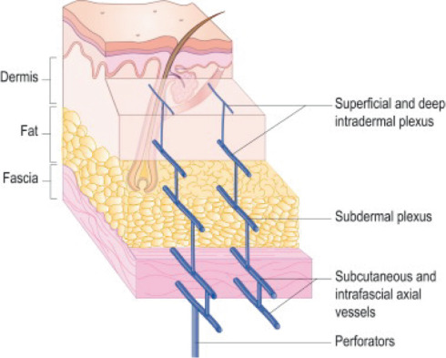
During the procedure, it is important to preserve the subdermal plexus ( Fig. 2.5.5 ). Subcutaneous fat or fascia may be included depending on the location and depth of the defect. Meticulous hemostasis before flap inset and skin closure is critical to prevent post-operative hematoma and possibly flap necrosis. Donor site defects may be closed primarily or, if undue tension precludes primary closure, covered with a skin graft.
Techniques
Rotation Flap
Principles
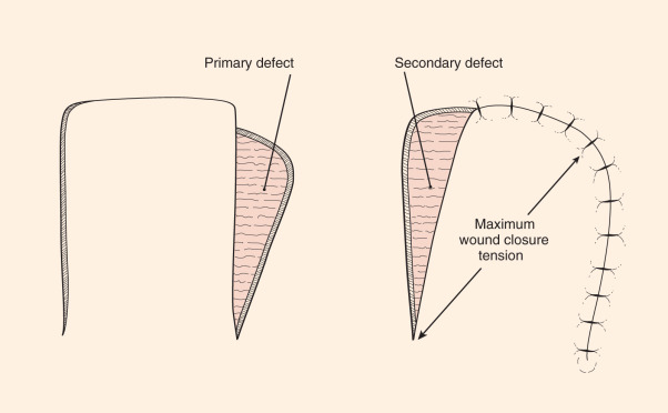
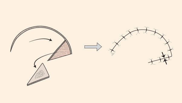
The rotation flap is commonly described as a semicircular flap that is rotated about a pivot point to close a triangular defect. Here the border of the defect becomes the leading edge of the flap with the remainder of the flap outline being drawn as the arc of a circle ( Fig. 2.5.6 ). The flap should be designed with the arc directed toward an area of tissue redundancy. The base of the flap, which is the radius of the large circle, should ideally be oriented inferiorly to allow for adequate lymphatic drainage post-operatively. Rotation flaps with a larger diameter and longer length require less rotation to fill a defect, and thus they result in less tension across the flap and suture lines. The curvilinear length of the flap should be at minimum four times the width of the defect.
Once raised, the flap is rotated to fill the defect, creating a length mismatch between the margin of the flap and the margin of the surrounding skin to which it will be sutured. This length discrepancy may be distributed along the length of the flap if there is sufficient skin laxity. At times, excessive outer skin contributes to the formation of a dog ear. This may be corrected by excision of a triangle of skin (Burow’s triangle) away from the flap. Hence, the flap base is not compromised and the length mismatch is corrected, which facilitates flap inset and skin closure ( Fig. 2.5.7A ).
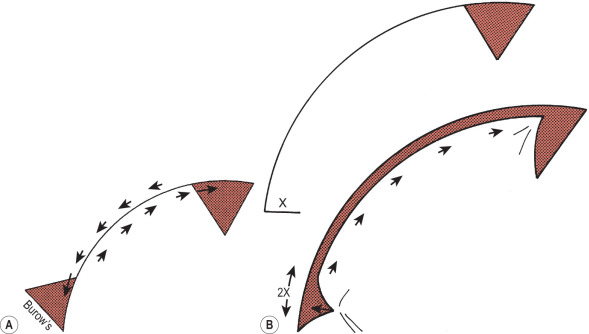
In general, the larger the flap in relation to the defect, the less tension will be on the final closure. A common mistake is to design the flap too small. This may then necessitate a back-cut toward the pivot point along the diameter of the semicircle farthest from the defect to decrease the tension across the suture line ( Fig. 2.5.7B ). Although tension is decreased by virtue of bringing the pivot point closer to the defect, it also decreases the width of the flap base, potentially compromising blood supply.
Clinical Examples
A rotation flap can cover a wide variety of defect sizes, making it a workhorse flap for various defects, including those of the scalp, face, and extremities as well as sacral pressure sores. Fig. 2.5.8 demonstrates the use of two rotational flaps to cover a scalp defect.
Transposition Flap
Principles
The transposition flap is commonly described as a rectangular flap that adjoins an existing defect and is moved laterally for defect coverage. In contrast to the rotation flap, the transposition flap is moved or transposed over an area of intact tissue as it is transferred into the defect. The more the flap is transposed, the shorter it becomes. As such, it is advisable to ensure that the flap extends beyond the defect ( Fig. 2.5.9 ). This design ensures that the flap is not too short after transfer and, thus, effectively prevents the need to use a back-cut in an attempt to decrease tension during flap inset. The donor site, which is commonly larger than the primary defect, is typically covered with a skin graft.