Key Words
transverse deficiency, congenital upper extremity, congenital amputation, symbrachydactyly, web space deepening, four-flap Z-plasty, local tissue rearrangement, distraction osteogenesis
Synopsis
Congenital transverse deficiency of the upper extremity is a rare but disabling condition in which all elements beyond a given level in the extremity are absent. The most common level is the proximal forearm, but it can occur at any level from the humerus to the metacarpals. This chapter describes the disorder, presentation, and an approach to management of patients with this condition. Surgery is not indicated in most transverse deficiencies at the forearm level or more proximal, but there are several surgical techniques that can improve function in patients with hand-level transverse deficiencies including symbrachydactyly. Described in detail is the technique of four-flap Z-plasty for first web space deepening, along with the postoperative care, common complications, and their management.
Clinical Problem
Congenital transverse deficiency is a rare but severe condition in which the upper extremity is truncated at a given level and all normal elements beyond this are absent. The estimated incidence is between 15 and 60 per 100,000 live births. In transverse deficiencies, a portion of the limb is missing, but the proximal supporting structures are intact, although typically hypoplastic. This is in contrast to longitudinal deficiency, wherein the proximal supporting structures are absent or severely deficient.
Historically, transverse deficiencies were often referred to as “congenital amputations,” but this is etiologically incorrect. As opposed to amniotic bands causing prenatal amputation of a limb that has already formed, transverse deficiency is considered a failure of formation of parts, type IA in the International Federation of Societies for Surgery of the Hand (IFSSH) system for congenital hand differences. The disorder is sporadic and is thought to be caused by disruption to the normal proximal-to-distal development of the extremity during embryological development of the upper limb.
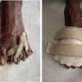
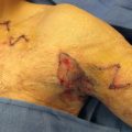
The management of transverse deficiencies requires an individualized approach, taking into account the level of the deficiency, current functional status, and the goals and desires of the child and family. In many cases throughout the world, surgical treatment is not indicated and children can adapt incredibly well to their differences with or without custom prosthetic devices. In particular, when the contralateral hand is unaffected, extensive reconstructive efforts are not usually indicated. In specific clinical situations, surgical interventions may be indicated to improve limb function. Examples include excision of nubbins or skin invaginations if causing secondary problems or interfering with prosthesis fit. In hand-level deficiencies, procedures that increase effective digital length may allow or improve prehensile function. Most commonly, this entails web space deepening with local flaps and tissue rearrangement techniques. Other procedures include skeletal lengthening using distraction osteogenesis, and vascularized or non-vascularized toe transfers. This chapter will provide an overview of transverse deficiency and surgical procedures to improve hand function, with a more in-depth look at web space deepening techniques.
Presentation
Patients with transverse deficiency present with congenital truncation of the upper limb, which may occur at any level from the humerus to the metacarpal. This may be detected on prenatal ultrasound examinations, or it may not be discovered until birth. Transverse deficiency can be differentiated from amniotic band syndrome (prenatal amputation), because osseous hypoplasia is a cardinal feature of the former but not the latter.
The left upper extremity is affected twice as often as the right, and males are affected more commonly than females. The most common level of truncation is at the proximal forearm, followed by the transcarpal, distal forearm, and distal humerus levels. Rudimentary finger “nubbins” are often present at the distal end of the truncated extremity, regardless of the level of deficiency. These often have nail plates and variably present extrinsic tendinous structures. Skin invaginations may also be present at the terminal end of the limb, which can cause issues with hygiene or infections.
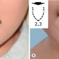
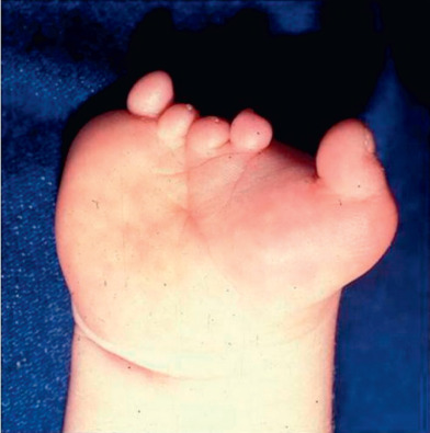
Symbrachydactyly, a condition characterized by shortened or absent fingers with various degrees of fusion, is now considered to be a type of transverse deficiency occurring at the level of the hand, although it is still classified as an “undergrowth” abnormality by IFSSH. Symbrachydactyly was classified into four types by Blauth and Gekeler. Type 1, short finger type, is characterized by four shortened, syndactylized fingers with an essentially normal thumb. Type 2, oligodactylic type, is characterized by central clefting or aplasia. Type 3, monodactylic type, demonstrates absence of all fingers with a normal thumb. Type 4, peromelic type, presents with an absence of all digits at the metacarpal level ( Fig. 5.4.1 ).
Etiology
One of the most important roles of the hand surgeon is to convey accurate information to the parents of a child born with congenital limb differences regarding the etiology of a condition, and to help to dispel feelings of guilt or parental responsibility that are commonly present. Culturally appropriate education is critical, especially when working in another country. Transverse deficiency is usually sporadic, and subsequent children are not thought to be at increased risk of limb differences.
There is an association with maternal intake of alcohol, tobacco, cocaine, or misoprostol, as well as riboflavin deficiency. There is also some evidence for an increased risk associated with chorionic villus sampling if the procedure occurs during the period of limb morphogenesis.
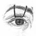
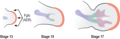
Nearly all congenital upper limb differences arise between gestational day 26, when the limb bud appears, and day 53, when the fingers are fully separated. The longitudinal development of the upper limb is driven by the apical ectodermal ridge (AER), a signaling center on the distal aspect of the limb bud that acts through fibroblast growth factors (FGFs) to render proximal to distal limb development and finally interdigital necrosis ( Fig. 5.4.2 ). The AER signals the differentiation of the underlying mesenchymal components of the limb bud, and is genetically programmed to induce appropriate differentiation through the time course of limb formation. A disruption of AER signaling during weeks 5 to 7 of gestation may lead to transverse deficiency. AER disruption is thought to be due to a vascular problem, either ischemia or bleeding, in most cases.
It is theorized that rudimentary digits, or nubbins, may be a result of a prenatal regenerative response to intrauterine trauma. Morphologically, these nubbins are similar to the distal portions of fingers, and can form on the end of limbs that are missing all other terminal structures. They can be found on the distal end of the limb, regardless of the level of truncation. It is theorized that either portions of the AER survive the insult and lead to the generation of the nubbins, or that the AER itself partially regenerates and induces the formation of nubbins.
Associated Conditions
Transverse deficiency usually presents unilaterally in otherwise healthy children without other congenital differences. Symbrachydactyly, however, may be associated with Poland syndrome, in which absence of the sternal head of pectoralis major and other muscles of the shoulder girdle may be absent or deficient.
Pre-Operative Management
Physical Examination and Key Anatomy
Transverse deficiencies should be characterized in terms of the level of truncation, and any associated distal rudimentary structures such as nubbins or invaginations. Furthermore, the function of adjacent joints such as the elbow and shoulder should be assessed and documented. In forearm-level transverse deficiencies, elbow flexion and extension are usually intact, but forearm rotation is commonly impaired by proximal radioulnar abnormalities. In patients with more distal transverse deficiencies at the hand or digit level, including those with symbrachydactyly, the functional usage of the hand should be determined. In particular, pinch and prehensile function should be assessed, because these functions may be restored or improved with surgical interventions. The best way to assess this in young children is to observe them playing with simple toys.
The fundamental function of the hand, prehensile function, can be achieved with a mobile thumb able to oppose other parts of the hand. In symbrachydactyly, the central rays are always affected, with variable involvement of the thumb and small finger. Depending on the specific morphology of the patient, web space deepening procedures can significantly improve prehensile function, particularly deepening of the first web space. Procedures requiring highly specialized equipment such as microsurgical instruments or distraction devices may not be possible or advisable in some facilities.
Pre-Operative Testing Needed
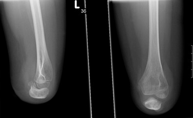
In forearm-level transverse deficiencies, standard x-rays of the elbow and forearm are generally the only diagnostic investigations required ( Fig. 5.4.3 ). Proximal radioulnar abnormalities are commonly present in forearm-level deficiencies. In patients with hand-level truncation, including symbrachydactyly, x-rays are useful to understand the underlying skeletal abnormalities and to assess whether certain interventions such as distraction osteogenesis may be possible for the patient. No specific pre-operative investigations are needed before web space deepening procedures, but close observation of the patient using the hand to play or manipulate objects will guide the surgeon in choosing the appropriate intervention, if any.