Chapter 20 Lipomyosculpture
• Morphological and histological analysis demonstrates differences in the superficial layer, regarding the layout of lobes, the shape disposition of interlobular septa tissue (collagen), and thickness of the different body parts.
• Marking before surgery is considered a key step for successful surgery. This should always be performed with the patient standing in front of the mirror. This ensures three-dimensional mobility and the patient can monitor all the surgeon’s movements and tracings.
• Ultrasonic energy (UAL) and vibroliposuction (VL) are useful as aids to conventional liposuction, but not for skin retraction.
• Liposuction must respect the histological peculiarities of the superficial layer of fat in order to stimulate skin retraction, and follow the muscle fibers’ direction; this makes it possible to carve the fat on the muscle, avoiding criss crossing.
Morpho-Histology of Subcutaneous Adipose Tissue
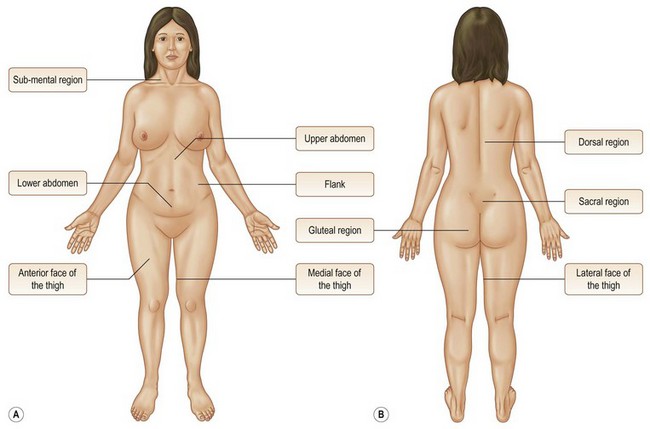
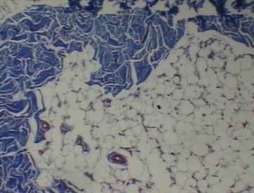
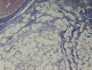
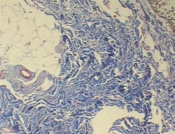
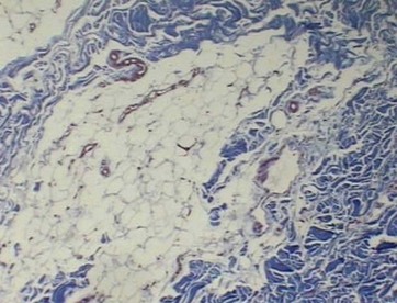
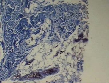
The subcutaneous tissue is divided into two layers. Its morphological and histological analysis demonstrates differences in the superficial layer, regarding the layout of lobes, shape, disposition of interlobular septa tissue (collagen), and thickness of different body parts (Fig. 20.1).
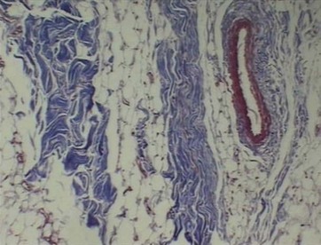
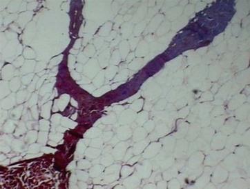
• Upper abdomen: irregular (50%), elliptical (40%) and rounded (10%) lobes with a parallel arrangement to the epidermis, thick (50%) and thin (50%) septa with a parallel arrangement in relation to the lobes (Fig. 20.2).
• Lower abdomen: irregular (50%), elliptical (30%) and rounded (20%) lobes with a parallel arrangement to the epidermis, thick (70%) and thin (30%) septa with a parallel arrangement in relation to the lobes (Fig. 20.3).
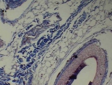
• Lateral thigh: irregular (80%), elliptical (10%) and rounded (10%) lobes with a parallel arrangement to the epidermis, thick (70%) and thin (30%) septa surrounding the lobes (Fig. 20.4).
• Anterior thigh: irregular (60%), elliptical (30%) and rounded (10%) lobes with a parallel arrangement to the epidermis, thick (50%) and thin (50%) septa surrounding and penetrating the lobes (Fig. 20.5).
• Medial thigh: irregular (90%) and elliptical (10%) lobes with a parallel and perpendicular arrangement to the epidermis, thick (30%) and thin (70%) septa surrounding and perforating the lobes (Fig. 20.6).
• Dorsal region: irregular (80%), elliptical (10%) and rounded (10%) lobes with a parallel arrangement to the epidermis, thick (80%) and thin (20%) septa (Fig. 20.7).
• Flank: irregular (70%), elliptical (20%) and rounded (10%) lobes with a perpendicular arrangement to the epidermis, thick (50%) and thin (50%) septa surrounding and perforating the lobes (Fig. 20.8).
• Gluteal region: irregular (80%), elliptical (10%) and rounded (10%) lobes with a parallel arrangement to the epidermis, thick (90%) and thin (10%) septa surrounding and perforating the lobes (Fig. 20.9).
< div class='tao-gold-member'>
Stay updated, free articles. Join our Telegram channel

Full access? Get Clinical Tree


