■ The lower back should be in a completely horizontal plane. This can be accomplished by lowering the leg portion of the OR table as much as possible to stretch the back and counter the natural convexity of the lower back.
■ Raising the leg extension with the knees flexed and lower legs horizontal/parallel to the floor will help to prevent the patient from slipping further down on the bed.
■ Flex arms at elbows, pad pressure points, and secure patient in place. Prep from midchest to just above buttocks.
PORT PLACEMENT
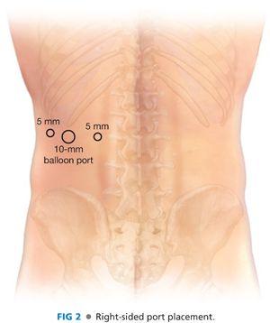
■ Three ports (lateral, middle, and medial) (FIG 2)
■ Medial: 5-mm “pediatric” or short port. Place this just lateral to paraspinous muscle and a few centimeters inferior to the costal margin.
■ Middle: 10-mm port with a “donut” balloon. Place this halfway in between the medial and lateral ports.
■ Lateral: 5-mm pediatric or short port. Place this as laterally as possible.

■ The middle port is placed first via a direct cutdown by making a transverse incision just large enough to admit a finger immediately inferior to the tip of 12th rib or the costal margin. The position of this port is determined as discussed earlier.
■ Dissection proceeds through the subcutaneous tissue to the muscle just inferior to the costal margin by spreading with a Metzenbaum scissors. The retroperitoneal space is entered by blunt dissection with the Metzenbaum scissors through the fascia and muscle using a technique similar to placing a chest tube (except that the hole is made directly inferior to the costal margin). Feeling the smooth undersurface of the ribs provides confirmation that the correct space has been entered. Use blunt dissection with the finger both laterally and medially to develop space for the placement of the subsequent 5-mm ports under direct palpation. Dissection should not be carried superiorly as this can make entering Gerota’s fascia more challenging.
■ Place the medial port 3 to 4 cm inferior to the costal margin just lateral to the paraspinous muscle entering the retroperitoneal space on a bias/angle cephalad of approximately 45 degrees such that the port enters the retroperitoneal space just inferior to the inferior margin of 12th rib. This angling of the port will improve visualization, obviating the need to torque the port.
■ Place the lateral port just inferior to the costal margin as far laterally as possible.
■ Place a balloon port in the middle port site to form a seal for the retroperitoneal insufflation.
DISSECTION OF THE RETROPERITONEAL SPACE
■ Insufflate to 20 mmHg, liberally increasing as needed to maximum of 30 mmHg.
■ Place camera into the middle port and a sealing device into the lateral port.
■ Enter Gerota’s fascia bluntly and open it widely from left to right.
■ Sweep the filmy posterior attachments of the periadrenal and perirenal fat anteriorly (i.e., toward the table). Continue this dissection to expose the peritoneum laterally, paraspinous muscle medially, and peritoneum superiorly.
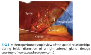
■ Switch camera to medial port and place a grasper into the middle port (FIG 3).

■ Identify the superior border of the kidney. Starting laterally, divide the adrenal-renal connections using a combination of blunt and sharp dissection. Carry this dissection toward the paraspinous muscle following the superior contour of the kidney, which will allow the kidney to be retracted inferomedially.
■
Stay updated, free articles. Join our Telegram channel

Full access? Get Clinical Tree


