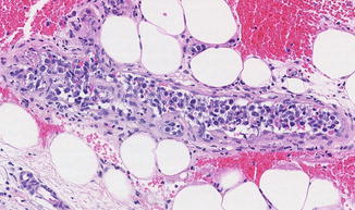
Fig. 69.1
IVBL: Dermal and subcutaneous vessels appear prominent in the scanning magnification

Fig. 69.2
IVBL: Subcutaneous vessel filled by large atypical lymphoid cells with nuclear pleomorphism

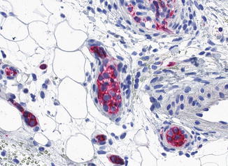
Fig. 69.3
IVBL: The intravascular tumor cells express the B-cell marker CD20
Differential Diagnosis
The diagnosis is based on the characteristic intravascular growth and the phenotype of the tumor cells. Among lymphomas, intravascular CD30+ anaplastic large T-cell lymphoma is distinguished by the phenotype of tumor cells. Benign atypical intravascular CD30+ T-cell proliferation due to trauma or inflammation represents a reactive condition mimicking intravascular lymphoma. Differential diagnosis also includes leukemia and intralymphatic histiocytosis as it can be seen after orthopedic metal implantation or in patients with rheumatoid arthritis.
Prognosis
IVBL carries an unfavorable prognosis. The cutaneous form displays however a better prognosis with 3-year survival rate of 56 % compared to the systemic form of IVBL (33 %).
Treatment
IVBL is treated with multiagent chemotherapy, which seems, however, not to be very effective.
Bibliography
Baum CL, Stone MS, Liu V. Atypical intravascular CD30+ T-cell proliferation following trauma in a healthy 17-year-old male: first reported case of a potential diagnostic pitfall and literature review. J Cutan Pathol. 2009;36:350–4.PubMedCrossRef
Stay updated, free articles. Join our Telegram channel

Full access? Get Clinical Tree


