(1)
Professor of Plastic Surgery, Director of Diabetic Wound Center, Director of Cell Therapy Laboratory, Korea University College of Medicine and Korea University Guro Hospital, Seoul, Republic of Korea (South Korea)
Abstract
The ideal dressing material should provide a moist environment to the wound while shielding it from bacterial invasion. The traditional gauze dressing cannot maintain a moisturized environment or absorb excess exudates, and gauze fibers are too widely spaced to effectively block out bacterial invasion. Recently, various interactive (or advanced) dressing materials have been developed to promote cell activity or to minimize scar formation and are currently in active use. One can choose from film, hydrocolloid, hydrogel, foam, hydrofiber, biologic, and composite dressings to suit one’s needs according to the condition of the wound. In this chapter, information of interactive wound dressings including classification, function, and selection is presented. In addition, advantages, action mechanisms, and precautions of silver- and iodine-based antimicrobial dressings are described. In particular, feature of a new polyurethane foam dressing impregnated with 3 % povidone-iodine, which has been recently developed to overcome the limitations of current silver-containing foam dressings, is demonstrated.
Keywords
Wound dressingInteractive dressingAntimicrobial dressingOverview
A wound dressing is an adjunct that is applied to a wound to promote healing and/or prevent further harm. A dressing is designed to be in direct contact with the wound. The ideal dressing material should provide a moist environment to the wound while shielding it from bacterial invasion, enabling the cells in the wound area to function in an active and stable manner. The most commonly used traditional dressing material – gauze – does not, in any way, promote cellular functions or activities. Gauze dressings cannot maintain a moisturized environment or absorb excess exudates, and gauze fibers are too widely spaced to effectively block out bacterial invasion. Recently, various dressing materials have been developed to promote cell activity or to minimize scar formation and are currently in active use. Over 3,000 dressing products have been registered. One can choose from film, hydrocolloid, hydrogel, foam, hydrofiber, biologic, and composite dressings to suit one’s needs according to the condition of the wound. In this chapter, information of interactive wound dressings including classification, function, and selection is presented.
Functions of Wound Dressings
A wound dressing should create a moist wound environment. If a wound is too wet, the dressing must absorb excess exudate. If a wound is too dry, the dressing should donate moisture to the wound bed. A moist wound environment facilitates all three phases of wound healing by trapping endogenously produced enzymes to facilitate autolytic debridement, preserve endogenously produced growth factors, and reduce patient pain complaints. A moist wound results in a more cosmetically appealing scar. A wound that is too wet can delay healing. If the surrounding skin is macerated, additional ulceration and infection can occur.
In addition, a wound dressing should provide thermal insulation (protecting the wound from temperature changes); provide a barrier to microorganisms to protect against infection; protect exposed nerves to decrease associated wound pain; eliminate dead space within the wound bed to prevent premature wound closure and abscess formation; remove debris, necrotic tissue, and foreign material; and finally provide adequate gas exchange between the wound and environment (Figs. 2.1, 2.2, and 2.3).

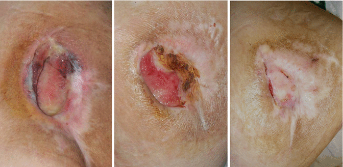
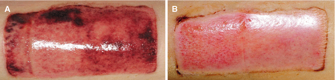
Fig. 2.1
Applying an appropriate wound dressing is essential to promote healing
Fig. 2.2
A palm-sized sacral sore wound was able to completely heal by using appropriate wound dressings only
Fig. 2.3
(A) A donor site wound of a split-thickness skin graft. (B) According to the selection of dressing materials, wound healing rates can vary. In this photo, a right side dressing induced faster healing than the left side dressing
Requisite Conditions for Dressing Materials
The ideal dressing material should provide a moist environment to the wound while shielding it from bacterial invasion, enabling the cells in the wound area to function in an active and stable manner. More specifically, in order to maintain adequate humidity, the surface of the dressing in contact with the wound should not dry out, while also absorbing excess exudate. The dressing should function as a barrier, protecting the wound from external bacterial invasions, and should be able to be removed both painlessly and atraumatically from the wound during dressing changes. Many studies have verified the fact that a moisturized environment promotes wound healing better than a dry one. Regenerating epithelial cells in a moist environment spread out smoothly along the wound surface, unlike those in a dry environment that selectively advance through the moist environment they can find – under the skin – leading to delayed and inefficient wound healing. In addition, polymorphonuclear (PMN) leukocytes, macrophages, proteinases, cellular growth factors, and other substances involved in wound healing are included in the wound exudate. In a dry environment, these substances would either be discharged externally or desiccated, rendering them useless, but a moist environment enables them to fulfill their functions, promoting more effective wound healing.
Wounds with a heavy bacterial load have an increased overall metabolic rate; this means that less oxygen and nutrients are distributed to normal cells, leading to decreased cellular function. Toxins and proteinases secreted from bacteria can also destroy cells and extracellular matrices, hence the need for dressing material to act as a barrier against external bacteria.
Interactive Dressing Materials
Judging by the aforementioned criteria for ideal dressing materials, the most commonly used traditional dressing material – gauze – does not, in any way, promote cellular functions or activities. Gauze dressings cannot maintain a moisturized environment or absorb excess exudates, and gauze fibers are too widely spaced to effectively block out bacterial invasion. Furthermore, regenerating wound tissues can grow into the gauze, effectively adhering the gauze to the wound; therefore, removing and changing such dressings can traumatize the wound, delaying wound healing and making dressing change a painful procedure. From this point of view, gauze dressings can only act as simple covers for wounds and do not functionally promote wound healing in any way (Fig. 2.4).
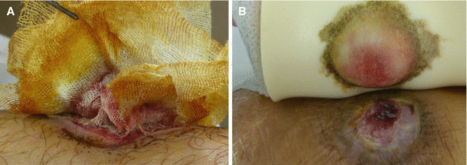
Fig. 2.4
(A) A gauze dressing adhering to the wound. (B) A foam dressing, one of the interactive dressing products. In comparison with a gauze dressing, a foam dressing has many advantages (details in Fig. 2.5)
Various dressing materials have been commercialized with the aim of supplementing the shortfalls of conventional gauze dressings. Over 3,000 dressing products have been registered. One can choose from film, hydrocolloid, hydrogel, foam, hydrofiber, biologic, composite, and antimicrobial dressings to suit one’s needs according to the condition of the wound.
Taking a closer look at the current most popular dressings among these dressing types, foam dressings have an outer protection layer with pores too small for bacteria to enter or exudates to ooze out through, while gases such as oxygen can be regularly exchanged through these same pores. The middle absorption layer was devised to keep and hold absorbed wound exudates, and the pores of the inner contact layer are of a size specifically devised to allow the passage of exudates but prevent ingrowth of regenerated tissue. These three layers functionally maintain a moist environment and protect the wound from bacterial invasion without adhering to the wound, enabling atraumatic and less painful dressing changes (Figs. 2.4 and 2.5).
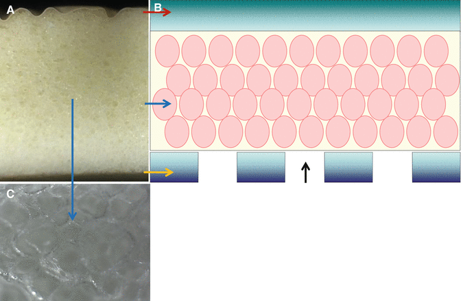
Fig. 2.5
(A) A magnifying view of the layers of a foam dressing. (B) The outer protective layer (a red arrow) prevents bacterial invasion. The middle absorptive layer (blue arrows) absorbs excessive exudates. The inner contact layer (a yellow arrow) prevents regenerating tissues from growing into (a black arrow) and adhering to the dressing. (C) Magnification of the holes in the middle layer (×100)
Classification
As mentioned, there are more than 3,000 wound dressing products available on the market today. They can be classified into 8 main categories according to the base of dressings.
Gauze and Impregnated Gauze Dressings
Traditionally, wound dressings were made from woven (cottons) or non-woven gauze (synthetic, more absorbent). Gauze with finer weave and smaller pores minimizes the risk of trauma to the wound bed. Gauze dressings are highly permeable and relatively nonocclusive, inexpensive, and used as a non-time or short-term use.
Impregnated gauzes are mesh gauze dressings impregnated with petrolatum, bismuth, or zinc. They are used as contact layers that function as secondary dressings which are nonadherent and increase the occlusiveness of standard gauze dressings.
Gauze dressings are commonly used for both infected and noninfected wounds of any size, shape, depth, or etiology. They are also the dressing of choice for very frequent dressing changes, infected wounds being treated with chemical agents, wounds requiring packing, and patients with fragile integument (roll gauze).
Impregnated gauzes can be used on granulating wound beds and burn wounds. They can also be used to prevent exposed tendon sheaths from dehydrating or adhering to dressings.
Precautions regarding use of gauze dressing include that woven gauze may require more force to remove and may leave residue or lint in the wound bed, causing formation of granulomas. If allowed to dehydrate, the dressing will adhere to the wound bed. Roll gauze should be applied snugly at an angle but without tension. Gauze dressings impregnated with bismuth or iodine are cytotoxic to inflammatory cells and may cause inflammatory response.
Films
Semipermeable film wound dressings are thin, flexible sheets of transparent polyurethane with adhesive backing. They are permeable to water vapor and gas, but impermeable to bacteria and water. They have little absorptive capabilities, but allow for visualization of the wound bed and conform to body contours.
Semipermeable film dressings can be commonly used on superficial wounds such as lacerations, abrasions, partial-thickness wounds, sutured wounds, and graft donor sites with minimal drainage. They may also be used on granular wounds and areas of friction.
To prevent maceration, a skin sealant must be applied. Film dressings should not be used on infected wounds, wounds with moderate to heavy drainage, or patients with fragile skin.
Hydrogels
Hydrogels are 8–99 % water- or glycerin-based wound dressings that are available in sheets, gels, or impregnated gauzes. They can only absorb a minimal amount of fluid, but donate moisture to dry wounds. Hydrogels are permeable to gas and water. Most hydrogels are almost nonadhesive, thus requiring a secondary dressing (Fig. 2.6).
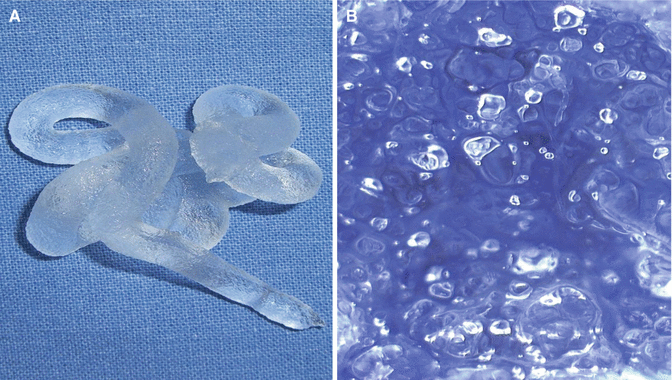
Fig. 2.6
(A) A hydrogel dressing. (B) Magnification (×100)
Hydrogels are indicated for any thickness wounds with minimal or moderate drainage. They can decrease pain and provide padding to decrease shear forces. They are also effective for softening eschars.
Hydrogels should not be used on infected wounds and on heavily draining wounds since they absorb fluids slowly.
Foams
Among the dressing types, foam dressings may be most commonly used and the current best seller, since they possess a number of important characteristics of ideal wound dressing based on the aforementioned criteria. Most of foam dressings are made of 3 layered polyurethane foams. An outer protection layer is hydrophobic with pores too small for bacteria to enter or exudates to ooze out, while permitting gases such as oxygen to be regularly exchanged. The middle absorption layer is devised to retain absorbed wound exudates. The inner contact layer is hydrophilic and has pores of a size specifically devised to allow the passage of exudates but prevent ingrowth of regenerated tissue. The 3 layers functionally maintain a moist environment and protect the wound from bacterial invasion without adhering to the wound, thus enabling atraumatic and less painful dressing changes. Foams are easy to apply and provide thermal insulation. Therefore, foam dressings can be universally used on wounds with minimal to heavy exudates.
Excessive wound exudate not only hinders the healing process, but also leads to maceration of the wound margins. Wound dressing should be able to quickly and effectively draw the exudates deeply into the absorbent material and reliably hold it there. Foam is an especially suitable dressing to accomplish these objectives.
Many foam dressings are primarily designed to absorb wound exudates and to provide soft cover to the wound site, in order to manage the wound environment. However, fluid absorption time, fluid absorption capacity, and fluid retention capacity are different according to individual foam dressings since they vary in compositions and modes of action. Particularly, the pores of foam dressings are designed to effectively absorb exudates, providing moist wound environment and decreased skin maceration. The pore size of a contact layer of foam dressings may have a significant impact on wound healing. Larger pores increase the growth of cells and tissue within the foam structure. The smaller the pore of the wound contact layer, the less likelihood of new healing wound tissue migration into the foam. Smaller pore size can also increase the absorption rate of wound exudates by capillary action. When the pores of the contact layer are placed in a wound bed, a concave meniscus is formed in pores. Adhesion occurs on pores drawing up exudate. The contact length between the top of the exudates and the pore is proportional to the diameter of the pore, while the weight of the exudates is proportional to the square of the pore diameter. Therefore, a small pore draws exudates higher than a larger pore. This causes exudates to be quickly drawn into the body of the foam and also ensures high retention for reliable exudate binding. Rapid absorption of exudates can prevent lateral spread of exudates to the periwound skin. The periwound skin requires protection from wound exudates to help prevent maceration and potential for further skin breakdown. The pore size of foam dressings varies widely according to products (25–500 μm).
Foam dressings are generally reserved for granulating wounds and skin graft donor sites (Figs. 2.7, 2.8, 2.9, and 2.10). Semipermeable foams are not indicated for dry or eschar-covered wounds, and skin sealant may be used to protect the periwound skin.
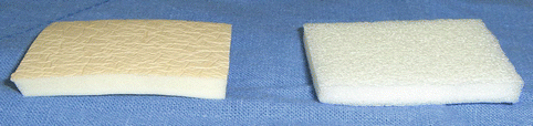
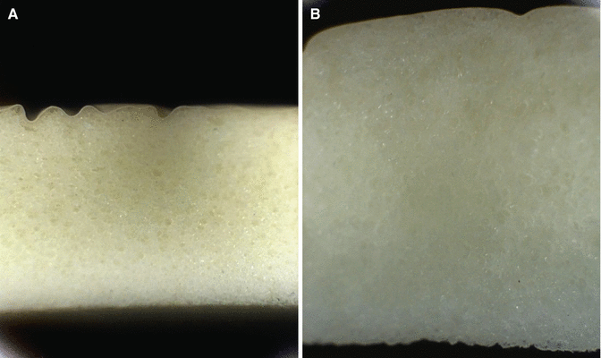
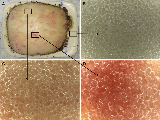
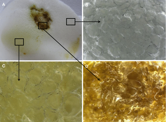
Fig. 2.7
Foam dressings with a backing film (left) and without a backing film (right)
Fig. 2.8
A foam dressing before (A) and after (B) absorbing exudates
Fig. 2.9
(A) The inner contact layer of a foam dressing backing with a protective film layer after absorbing exudates. Magnifications (×100) of the layer with no exudates (B), serous exudates (C), and sanguineous exudates (D)
Fig. 2.10
(A) The inner contact layer of a foam dressing without a back film layer after absorbing exudates. Magnifications (×100) of the layer with no (B), serous (C), and sanguineous (D) exudates. Unlike the foam dressing with an outer film layer, the dressing cannot maintain moisture
Hydrocolloids
Hydrocolloids contain hydrophilic colloidal particles such as gelatin, pectin, and cellulose with a strong film or a foam adhesive backing sheet. They are impermeable to water, gas, and bacteria and therefore can be effective barriers against urine, stool, and pathogenic microorganisms. They absorb fluids slowly by swelling into a gel-like mass. Hydrocolloids often leave residues after removal. Furthermore, they provide thermal insulation (Figs. 2.11 and 2.12).
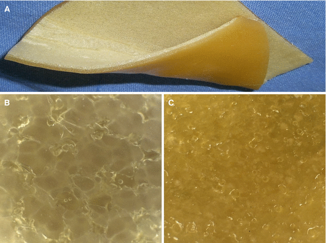
Fig. 2.11


(A) A hydrocolloid dressing. Magnifications (×100) of the outer (B) and the inner (C) layers
Related posts:
Stay updated, free articles. Join our Telegram channel
Full access? Get Clinical Tree
