(1)
Professor of Plastic Surgery, Director of Diabetic Wound Center, Director of Cell Therapy Laboratory, Korea University College of Medicine and Korea University Guro Hospital, Seoul, Republic of Korea (South Korea)
Abstract
We all experience our fair share of wounds during the course of our lives. We get pricked by thorns, scratched by sharp objects, sunburned at the beach, and scalded by hot water. Accidents can lead to our skin being peeled or sliced off, and many of us may undergo surgical procedures which inevitably result in surgical wounds on our bodies. Selecting an appropriate wound healing strategy is crucial for successful wound healing in that it can minimize the risk of complications, enhance the speed of wound healing, and minimize scar formation after the wound has fully healed. During the past few decades, various technologies have been developed for optimal wound healing. In order to understand new techniques, procedure, and materials in wound healing, medical professionals should have a basic knowledge of wound healing. In this chapter, clinically useful anatomy of the skin, terminology and documentation for wounds, basic wound healing process, and conventional wound healing methods will be briefly described prior to the main topics of this book.
Keywords
AnatomySkinWound healingClinical Anatomy of the Skin
Our skin layer has many crucial functions, but the main role is barrier function, that is, protecting our body from external elements. Unless the wound is properly healed, the integrity of the skin barrier is compromised and allows external germs such as bacteria and viruses to freely invade our body. This can even lead to critical organ damage. Moreover, the skin layer comprises an important aspect of one’s appearance, which of course has a significant influence on one’s social life; thus, the skin layer can also be regarded as an important factor of mental health.
Since the skin is such a critical element of both physical and mental health, optimal care must be given to any sustained wounds, so that the damaged skin region is restored to a structure and form similar to the original as quickly as possible for the skin to resume its functions. In order to give such optimal wound care, one must first understand the anatomy of the skin. The skin is the largest organ of the body. It weighs in the range of 2.7–3.6 kg and receives 1/3 of the body’s blood volume. The thickness of the skin varies from 0.5 to 6.0 mm. The skin consists of cells and extracellular matrices. There are three layers in the skin. The epidermis is the thin, outer layer of the skin. The dermis is a thicker, inner layer. The subcutaneous fatty tissue (hypodermis) is a layer of loose connective tissue lying beneath the dermis (Fig. 1.1).
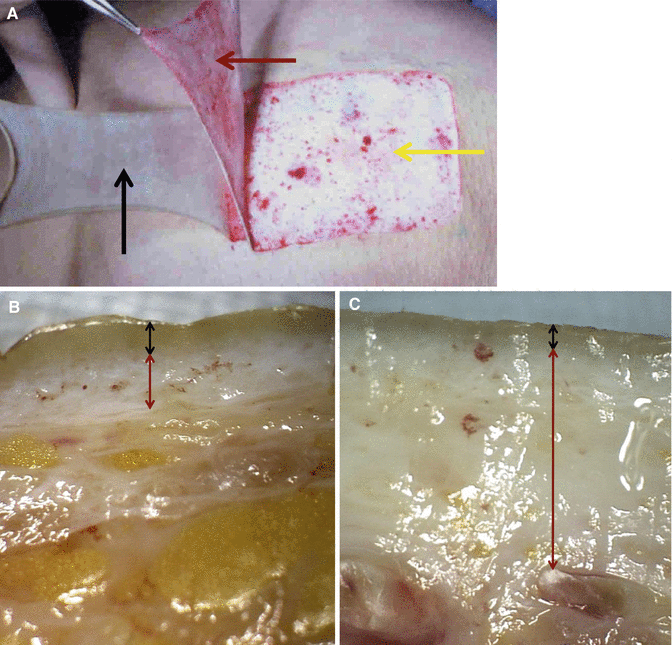
Fig. 1.1
Skin layers. (A) Epidermis (a black arrow), dermis (a red arrow), and hypodermis (a yellow arrow). (B) Epidermis (a black arrow) and dermis (a red arrow) of the thin skin (hand dorsum). (C) Epidermis (a black arrow) and dermis (a red arrow) of the thick skin (palm skin)
Epidermis
The thickness of the epidermis varies in different types of skin. It is the thinnest on the eyelids at 0.05 mm and the thickest on the palms and soles at 1.5 mm. The epidermis is an avascular layer receiving blood supply from the dermis across the semipermeable basement membrane.
Epidermal Layers
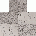
The epidermis contains five layers. From bottom to top, the layers are named stratum basale, stratum spinosum, stratum granulosum, stratum lucidum, and stratum corneum. The thickness and layers of the epidermis vary depending on the location of the body (Figs. 1.2 and 1.3).
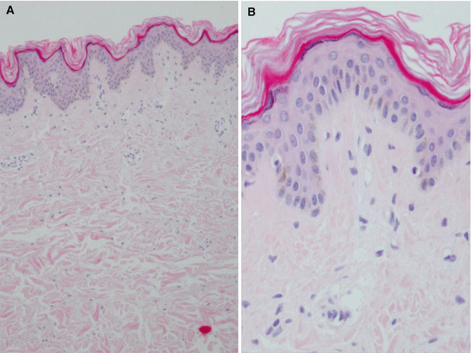
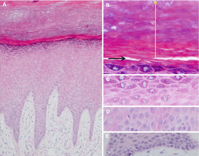
Fig. 1.2
Epidermis of the thigh. The number of epidermal layer is small. (A) ×100. (B) ×400
Fig. 1.3
Epidermis of the plantar skin. In comparison with the thigh skin (Fig. 1.2), there are much more layers in the epidermis. (A) ×100. (B) ×400. Horny (a yellow arrow) and lucid (a black arrow) layers. (C) Granular layer. ×400. (D) Spinous layer. ×400. (E) Basal layer. ×400
The stratum basale (basal layer) is the deepest layer with a thickness of a single cell. It is the only layer of the epidermis in which cells undergo mitosis. The stratum basale forms the dermal-epidermal junction (basement membrane zone), which separates the epidermis from the dermis. The stratum spinosum (spinous layer) consists of several rows of more mature keratinocytes, which appear spiny under a microscope. The stratum granulosum (granular layer) contains 3–5 flattened cell rows comprising a higher concentration of keratin. The stratum lucidum (lucid layer) is a thin, clear layer of dead skin cells found in the thick skin such as the palms and soles. The stratum corneum (horny layer) consists of dead cells (corneocytes) and keratin. This layer prevents water evaporation, absorbs water, and easily sheds itself.
Epidermal Cells
The keratinocytes are major cells of the epidermis, making up approximately 90 % of epidermal cells. These cells are responsible for the toughness of the skin. They produce keratin and form the basic component of hair, skin, and nails. Langerhans cells protect the body against infection by attacking and engulfing foreign materials. Melanocytes are responsible for producing melanin (Fig. 1.4). While the number of melanocytes remains the same in individual body regions in all human beings, their activity can vary between different body regions and across individuals. In white and oriental skin, the melanosomes are packed in “aggregates,” but in black skin, they are larger and distributed more evenly. The number of melanosomes in the keratinocytes increases with ultraviolet (UV) radiation exposure, while their distribution remains largely unaffected. Merkel cells are mechanoreceptors that provide information on light touch sensation.
Fig. 1.4
Melanocytes and melanin (brown color)
Epidermal Appendages
The sebaceous glands secrete sebum into hair follicles. Sebum is an oily substance, which provides moisture and softness to the skin. Sebum also acts as a barrier against foreign substances (Fig. 1.5). Hair contributes to the appearance, body temperature, protection, and sensation. The eccrine sweat glands produce sweat to help regulate body temperature and assist with elimination of waste products. The apocrine sweat glands produce sweat, which is responsible for “body odor.” Nails are made of dead cells that contain keratin.
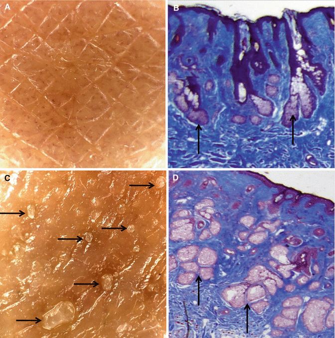
Fig. 1.5
Differences can be observed in the development of the sebaceous glands according to each individual. (A, B) Skin with less developed sebaceous glands (arrows). (C, D) Skin with well-developed sebaceous glands (arrows)
Functions of the Epidermis
The primary function of the epidermis is to act as a protective barrier against the outside environment. The acidic coating protects the skin from microorganisms. The tough keratin layer protects the body from invasion and infection and helps to seal in moisture. Langerhans cells provide allergen recognition and assist with immunity. The ability of the skin to hold water is primarily due to the stratum corneum and is critical for maintaining healthy skin. The amount and distribution of melanin pigment in the epidermis is the main reason for variation in skin color. The epidermis is a contributing factor in self-image since skin, hair, and nails originate in the epidermis. Vitamin D is synthesized in the epidermis upon exposure to UV-B radiation, primarily in keratinocytes of the stratum basale and stratum spinosum layers of the epidermis.
Dermis
The dermis varies in thickness depending on the location of the skin. It is 0.3–0.5 mm on the eyelid and 3.0–6.0 mm on the back.
Dermal Layers
The two layers of the dermis are the papillary and reticular layers. The papillary dermis is an upper layer, named for its fingerlike projections called papillae. The reticular dermis is a lower layer, which is denser than the papillary dermis. This layer gives strength to the skin.
Dermal Cells
Fibroblasts are the main cells of the dermis, which produce collagen, elastin, granulation tissue, and cytokines including growth factors. Macrophages and the white blood cells help fight infection. Mast cells help initiate inflammation through secretion of histamine, enzymes, and chemical mediators.
Dermal Components
Collagen is a protein that gives strength to the dermis. Elastin is a protein that makes the skin pliable. Blood vessels supply nutrients and oxygen to the skin and take away cell waste (Fig. 1.6). Lymph vessels transport lymph, a fluid that contains the infection-fighting cells of the immune system, and remove excess proteins in the interstitial tissues. Nerve endings of pain and touch receptors called Meissner corpuscles transmit sensations of pain, itch, pressure, and temperature.
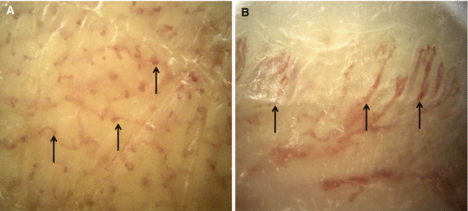
Fig. 1.6
Subdermal vascular plexus supplying nutrients and oxygen to the skin (arrows, ×100). (A) Surface of the skin. (B) Cross section of the skin
Functions of the Dermis
The dermis houses epidermal appendages. Infection is controlled by many cells located within the dermis. The dermis also provides nutritional support for itself and the epidermis, thermoregulation through the superficial vasculature, and sensation through the nerve receptors in the dermis.
Subcutaneous Tissue
The subcutaneous tissue is a layer of fat and connective tissue that houses larger blood vessels and nerves. This layer is important in the regulation of temperature of the skin itself and the body. The size of this layer varies throughout the body and from person to person.
Wound Overview
Definition
The term “wound” refers to a condition in which the normal skin structure is broken or destroyed, though the severity and depth may vary widely. Wounds include not only tears or lacerations in the skin layer with exposure of the subcutaneous tissue (an open wound) but also contusion from blunt objects (a closed wound). However, in pathology, the term is confined to cases in which the trauma sustained by skin structures has penetrated through the epidermis and inflicted damage on the dermis.
Classification
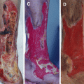
Wound Surface
The types of open wounds are incisions, lacerations, abrasions, puncture wounds, penetration wounds, and through-and-through wounds. The types of closed wounds are contusions (bruises), hematomas, and crush injury. Closed wounds have fewer categories but are just as dangerous as open wounds (Fig. 1.7).
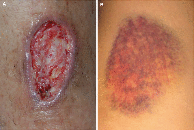
Fig. 1.7
Open (A) and closed (B) wounds
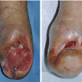
Chronicity
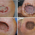
Acute wounds are those that heal within a predictable amount of time; in general 2 weeks in a healthy person for most wounds (Fig. 1.8). Chronic wounds are wounds that fail to heal properly or are in slow or stagnated healing; wounds that do not show signs of healing within 3–6 weeks are often considered chronic. The reason for chronic wounds can be due to many things such as diabetes, autoimmune diseases, chemical agents, radiotherapeutic agents, infections, and peripheral vascular diseases (Fig. 1.9).
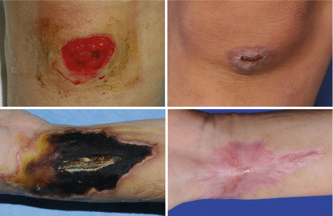
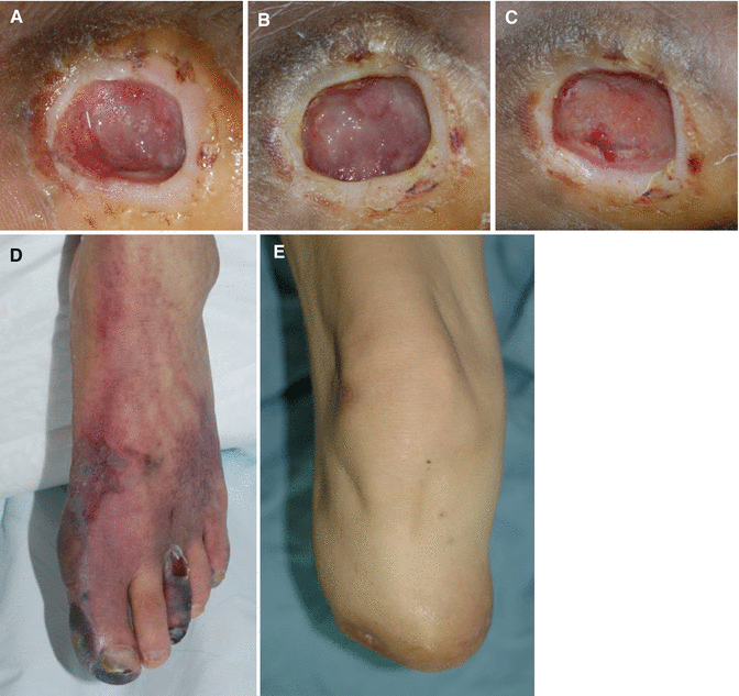
Fig. 1.8
Acute wounds. Acute wounds heal within a predictable amount of time
Fig. 1.9
Chronic wounds. This wound (A) does not show any sign of healing after 1 month (B) and 2 months (C). (D, E) Toe chronic wounds were finally closed by below-knee amputation
According to Depth
Superficial wounds affect only the epidermis. Partial-thickness wounds involve the epidermis and part of the dermis. Full-thickness wounds extend through the epidermis and dermis. Full-thickness wounds may extend into the subcutaneous tissue, fascia, and muscle.
Wound Documentation
Basic Examination
Patient history and systemic review should be recorded. Patient history includes demographics, lifestyle, and general medical history. Purposes of the systemic review are to identify the risk factors and underlying disease processes.
Wound Types
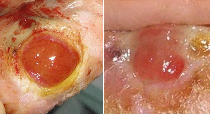
Granulation Tissue
Epithelialization
Epithelialization can appear deep pink in color, then progress to pearly pink and again to light purple from the edges in full-thickness wounds or may form islands in the wound base in superficial wounds (Figs. 1.12, 1.13, and 1.14).
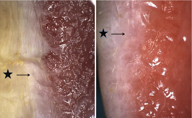
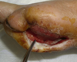
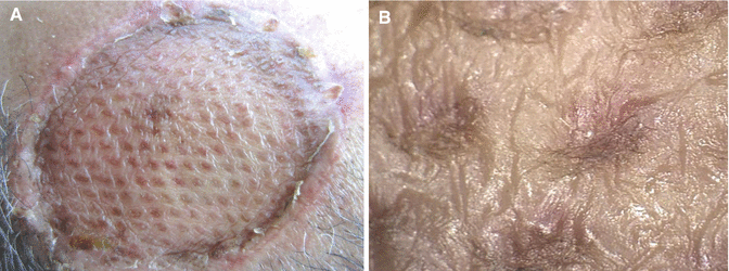
Fig. 1.12
Epithelialization (arrows) appears from the edges of the wounds (stars)
Fig. 1.13
Unfavorable epithelialization, which progresses inside of the wound bed
Fig. 1.14
(A) Epithelialization in a meshed skin graft. (B) A magnifying view
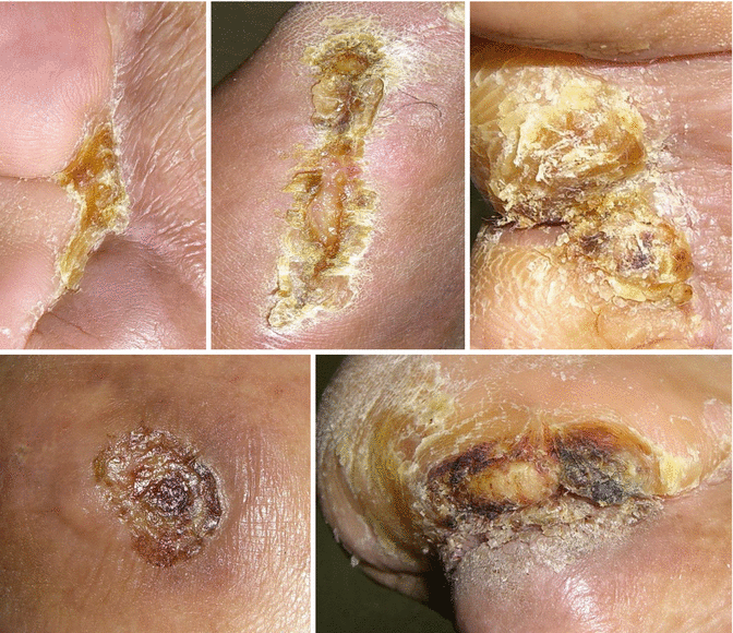
Necrotic Wound
Necrotic wounds are localized defects or excavation of the skin or underlying soft tissue that contains dead, avascular tissue. The level and type of tissue death influences the clinical appearance of the necrotic tissue. Examples of necrosis include white/gray nonviable tissue, stringy, yellow or tan sloughs, and hard black eschars (Fig. 1.15).
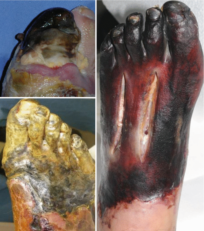
Fig. 1.15
Necrotic wounds
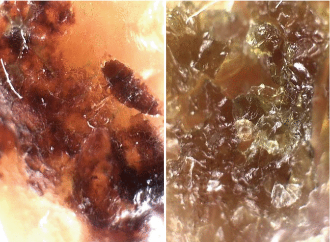
Eschar
An eschar is a hardened dry crust of necrotic tissue that may form over a wound. It is usually thick, leathery, and black. A white eschar indicates total ischemia (deficiency of blood) of the tissue. A red (or brown) eschar indicates hemoglobin from destroyed red blood cells (Figs. 1.16, 1.17, and 1.18).
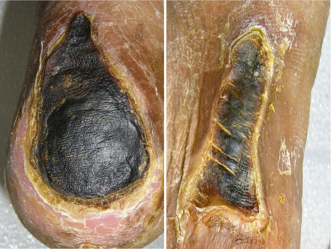
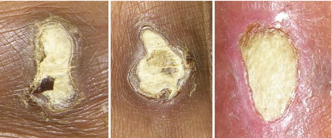
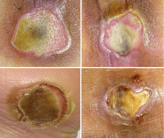
Fig. 1.16
Black eschars
Fig. 1.17
White eschars
Fig. 1.18
Brown eschars
Scab
A scab is a collection of dried blood and serum over a wound which has formed during the wound healing process. It is a combination of platelets, red blood cells, white blood cells, fibrin, and plasma. As the combination dries out, the scabs usually take on a deep, rusty brown color and develop crusty edges. Scabs generally remain firmly in place until the skin underneath is repaired and new skin cells appear.
Scabs actually prevent new skin cells from forming, which can result in longer healing time. Preventing scabs is the best way to promote healing. However, removing scabs is also dangerous since scabs function as protective caps over the wound and prevent dirt, germs, and other contaminants from entering the wound bed. In addition, if scabs are prematurely removed, the revealed skin could be red and oozing. New scabs may reform, but often, the new skin develops scar tissue.
Crust
Crust means any hard outer portion or surface area of solid matter. When a wound is described, a crust is used as a generic term describing an eschar and a scab. Generally, however, a crust over a wound is called a scab.
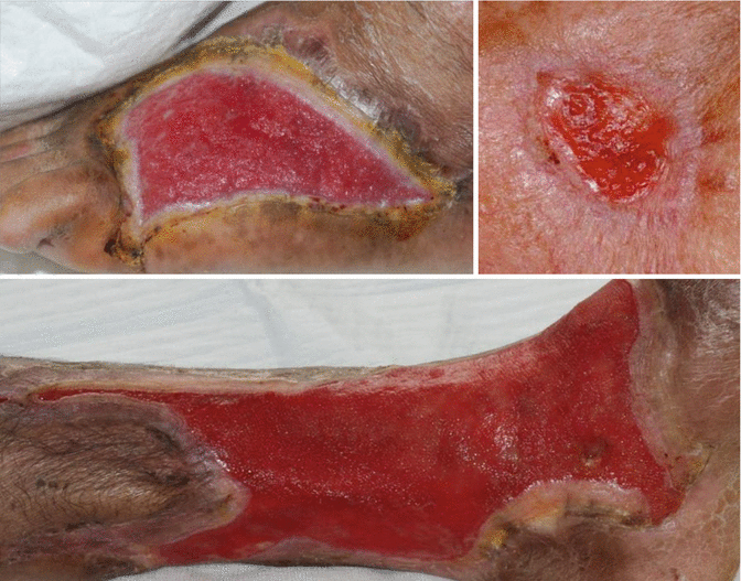
Slough
A slough is a layer or mass of dead tissue separated from the surrounding living tissue in a wound (Figs. 1.21, 1.22, and 1.23).
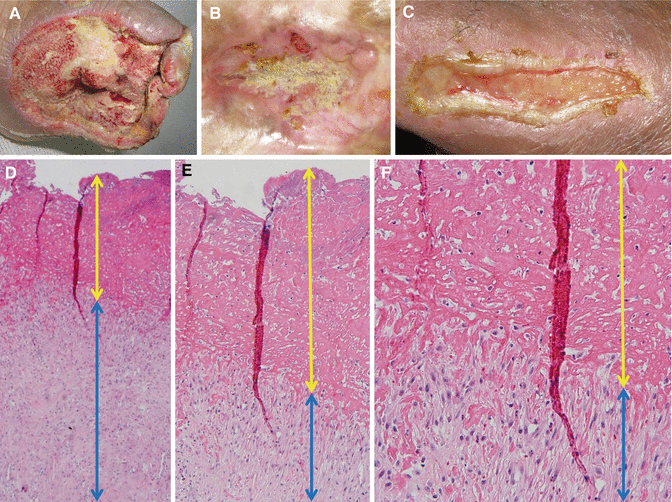
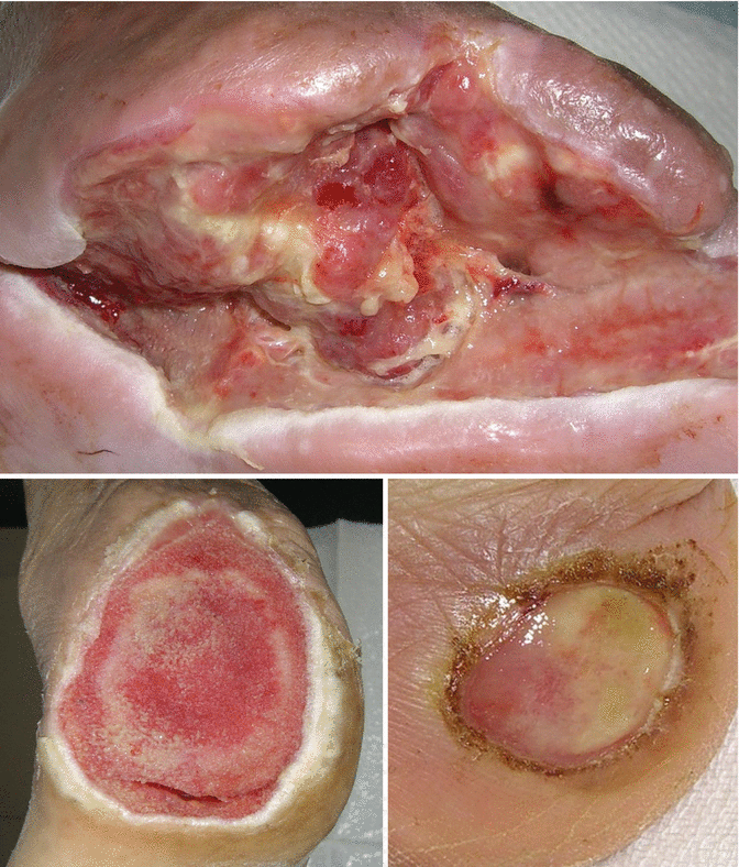
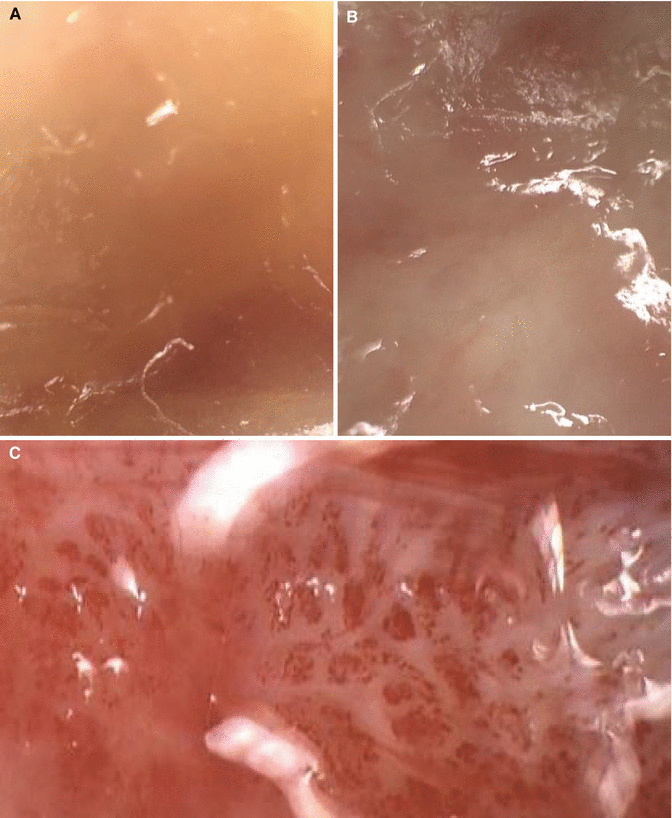
Fig. 1.21
Sloughs. (A–C) Gross findings. (D–F) Microscopic views (×40, ×100, and ×200, respectively). Yellow and blue arrows indicate slough and granulation tissue layers, respectively
Fig. 1.22
Mixed wounds having granulations and sloughs
Fig. 1.23
(A, B) Close-up views of sloughs over granulation. (C) After removal of the slough, healthy granulation tissue appeared
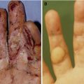
Callus (Callosity)
A callus is an especially toughened area of the skin which has become relatively thick and hard in response to repeated friction, pressure, or other irritations (Fig. 1.24).
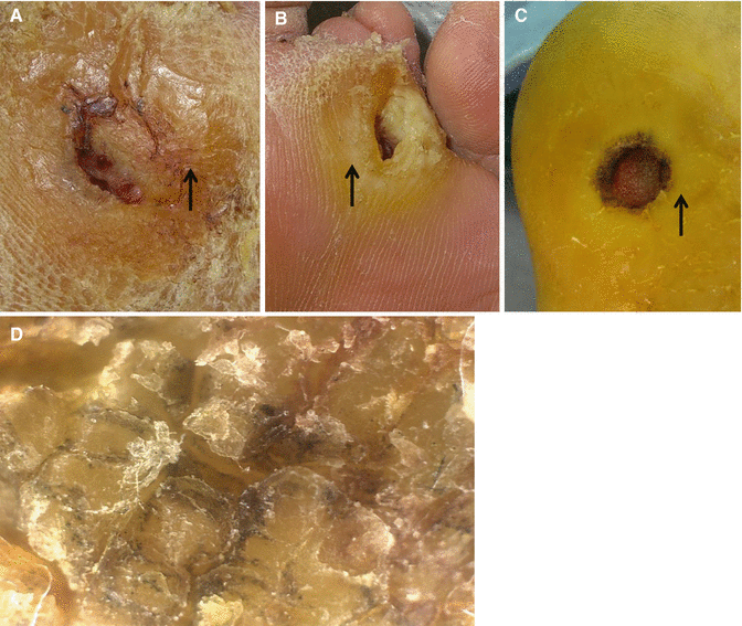
Fig. 1.24
Calluses (A–C, arrows) and a close-up view (D)
Scale
A scale is a thin piece of keratin layer of the skin that is produced because of abnormal skin conditions, most frequently excessive dryness (Figs. 1.25 and 1.26).

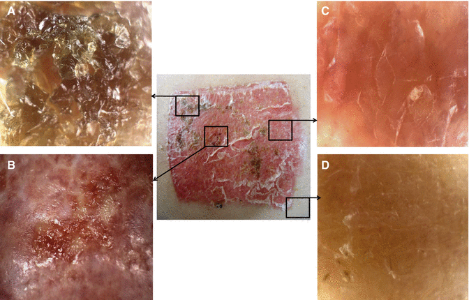
Fig. 1.25
Scales (A–C) and a close-up view (D)
Fig. 1.26
A donor site of split-thickness skin graft showing several wound types. (A) Scab or crust. (B) Unhealed site. (C) Epithelization with scale. (D) Normal skin
Wound Characteristics
Location and size of the wound should be recorded. Presence of sinus tract or undermining should also be examined. Sinus tract (tunneling) is a course or pathway that can extend into any direction from the wound through tissue and/or bone, resulting in dead space. Undermining is tissue destruction underlying the intact skin along the wound margins. Wound edges should be carefully observed: healing (evidence of epithelialization), rolled, or callused. Wound drainage should be documented focusing on type (serous, sanguinous, serosanguinous, or purulent), amount (none, minimal, moderate, or copious), and odor (strong, foul, pungent, fecal, musty, or sweet). Condition of the periwound is additionally examined: hydration/skin turgor, color, maceration, edema, induration, pitting, or temperature.
Related posts:
Stay updated, free articles. Join our Telegram channel

Full access? Get Clinical Tree


