(1)
Professor of Plastic Surgery, Director of Diabetic Wound Center, Director of Cell Therapy Laboratory, Korea University College of Medicine and Korea University Guro Hospital, Seoul, Republic of Korea (South Korea)
Abstract
In planning reconstruction for wounds, efforts should include choosing the safest and least invasive method with a goal of achieving optimal functional and cosmetic outcomes. Although a variety of methods can be used to cover defects, skin grafting is generally straightforward with a relatively low risk of complications. However, the two major concerns of skin grafting are poor matching of colors in the recipient site and donor site morbidity. To minimize the limitations of the classic skin graft, the author has developed an autogenous dermis graft, which is a deepithelialized skin graft, and has reported promising results on the coverage of small- to medium-sized wounds. The important aspects of this method include the immediate return of the epidermis to the donor site thereby overcoming donor site morbidity and minimizing pigment mismatch between the graft and the surrounding skin by restoring the epidermal portion of the recipient site through inducing epithelialization from the adjacent skin. To make the autogenous dermis graft even easier, the author has also used allogenic and artificial dermis for surface grafts. Based on the author’s experience, allogenic dermis is difficult to be taken by the wound bed. In addition, resultant scar after wound healing is usually not satisfactory. In the case of artificial dermis, clinical results are generally acceptable. Collagen sponge and hyaluronic acid sheets are two main commercial materials that are frequently used. In this chapter, the author presents the reliability of these biologic dermis grafts for wound coverage.
Keywords
Skin graftDermis graftAllogenic dermisArtificial dermisProper selection of the reconstruction method for the skin and soft tissue defect is the key to achieving successful results, especially in cases involving the face. For example, surgical excision of skin cancer on the face leaves a skin and soft tissue defect which requires reconstruction (Fig. 3.1). In planning reconstruction for wounds, efforts should include choosing the safest and least invasive method with a goal of achieving optimal functional and cosmetic outcomes. A variety of methods can be used to cover defects, such as healing by secondary intention, primary closures, skin grafts, and local flaps.

Fig. 3.1
Because the face is the most common site of skin cancer, cosmetic outcomes as well as complete cures should be considered in the selection of a treatment modality. (A–C) A skin and soft tissue defect created by resection of a basal cell carcinoma on the face
Healing by secondary intention usually leaves significantly conspicuous and unfavorable scars. Darker skin is more prone to hypertrophic scarring or keloid formation. The role of primary closure is limited because of the size and shape of the defect. Only small defects with elliptical shapes yield satisfactory results after primary closure. A local flap is usually considered the treatment of choice in covering a small facial defect because it may provide several advantages, such as decreased scar contracture and satisfactory contour, color, and texture matching. In some cases, however, a local flap is just not feasible, mainly due to the limitation of the size and arc of rotation, particularly in young patients. Poor flap design and inappropriate incisions may contribute to unacceptable scars, violation of aesthetic subunits, and resultant disfigurement on difficult-to-treat areas. In addition, a compromise in flap vascularity and inadequate wound closure created by insufficient undermining, lack of deep closure, excessive tension, or inadequate approximation of wound edges are predisposing factors to unfavorable results.
Skin grafting is generally straightforward with a relatively low risk of complications. Skin grafting is the easiest and the most reliable way to obtain proper wound coverage. However, the two major concerns of skin grafting are poor matching of colors in the recipient site and donor site morbidity including pain, discomfort, and hypertrophic scarring. Major pigment mismatches are common in split-thickness grafts particularly in darker-skinned patients including Asians. Several factors may play a role in the color mismatch of a regular skin graft. These include the amount of melanin, the degree of transfer of melanosomes to keratinocytes, and the number of melanocytes. Because melanocytes are localized to the basal cell layer of the epidermis, the origin of such color mismatch has been assumed to lie in the epidermal melanin. Because regular skin grafts that are transferred to new locations maintain their epidermal specificity, a pigmentation difference with the surrounding skin is unavoidable.
To minimize the limitations of the classic skin graft, the author has used autogenous, allogenic, and artificial dermis grafts for small- to medium-sized wounds. In this chapter, the author presents the reliability of biologic dermis grafts for wound coverage.
Autogenous Dermis Graft
The author has developed an autogenous dermis graft, which is a deepithelialized skin graft, and has reported promising results on the coverage of small- to medium-sized wounds on the body. The important aspects of this method include the immediate return of the epidermis to the donor site thereby overcoming donor site morbidity and minimizing pigment mismatch between the graft and the surrounding skin by restoring the epidermal portion of the recipient site through inducing epithelialization from the adjacent skin.
Surgical Technique
After sharp debridement of the recipient site, the skin and underlying subcutaneous tissue of the wound margin are undermined approximately 5 mm in length along the periphery. After meticulous hemostasis, the defect size of the recipient site is measured. A thin epidermal flap is elevated on the gluteal area or the lateral thigh using a Zimmer dermatome, in which the thickness is set to 0.010 in. The blade is then reset to 0.016–0.020 in., and the dermis to be grafted is cut from the same area. The previously elevated epidermal flap is replaced on the donor site and sutured with Prolene. The harvested dermis is tailored to the size and shape of the defect including the undermined area and is transferred to the recipient site. The edge of the dermis graft is inserted into the undermined wound margin and fixed to the wound bed along the circumference of the defect using PDS sutures. The undermined marginal skin over the grafted dermis is then fixed to the underlying dermis using Prolene sutures (Fig. 3.2). The skin sutures are removed after approximately 5–7 days in order to minimize the stitch marks. After wound healing, sunlight is avoided for the first 3–6 months using sun-blocking agents.
Fig. 3.2
Autogenous dermis grafting. (A) The thin epidermal flap is elevated from the donor site, and the dermis to be grafted is then cut on the same area. (B) The elevated epidermal flap is replaced on the donor site. (C) The edge of the dermis graft is inserted into the undermined wound margin and fixed to the wound bed along the circumference of the defect
Indication
Dermis grafts are indicated in any cases of small- to medium-sized skin defects (smaller than 50 cm2 based on the author’s experience) that are candidates for regular skin grafting. In particular, a dermis graft is likely to have its greatest reconstructive role on exposed areas such as the face, neck, forearms, and hands. In fairly large wounds, the return of patients to their normal daily activities is likely to be delayed because the epithelialization of the graft takes a longer time. There is no data on the critical size or thickness of the dermis that can produce scar-free wound healing. Further research may be needed on this issue.
A full-thickness small-sized wound, which is left to heal by secondary intention, sometimes results in acceptable scarring in the concave area of the skin. In some wounds, however, a period of several weeks or months is required for complete healing, particularly in actinically damaged, fragile skin. Wounds on convex areas of the skin heal rather poorly (depressed scar) via secondary intention. The resultant wound contraction, which is an important clinical component, is not an uncommon problem.
Controlled Clinical Study
The author has evaluated the efficacy of the dermis graft used for wound coverage by comparing it with that of a regular skin graft.
An autogenous dermis graft was applied to 53 patients, ranging in age from 6 to 61 years (average, 36.5 years). The size of the wound ranged from 2.7 to 42.0 cm2. The recipient sites included the face, forearm, hands, lower extremities, and back. A regular split-thickness skin graft procedure was simultaneously performed on the wounds of approximately the same size and location as the dermis graft in 33 patients. The size of the regular skin graft wound ranged from 2.3 to 42.0 cm2.
The graft take was complete in all patients in both groups. Refilling of the blood vessels and multiple pinpoint bleeding were observed on the grafted dermis within the first 2–3 days. Reepithelialization occurred progressively from the periphery to the center of the grafted dermis.
The whole wound of the dermis graft reepithelialized after grafting within 11–20 days (average 15.5 ± 1.9 days). The skin-grafted wounds were healed by 7–16 days (average 11.8 ± 1.6 days). In the long-term follow-up, good quality skin characteristics were achieved in the dermis graft. A comparison of the scars at the recipient site showed that the dermis graft was superior to the regular skin graft in terms of pigmentation, height, and vascularity. No significant differences in pliability were detected. Patient satisfaction in the dermis graft group was also better than that in the skin graft group. Regarding the donor sites, the results of the dermis graft were also satisfactory. All the donor sites of the dermis graft healed within 9 days (average 7.5 ± 0.8 days). In contrast, those of the regular skin graft required 11–16 days to heal (average 12.8 ± 1.1 days). Moreover, the donor sites of the dermis graft were superior to those of the skin graft in terms of scar quality and patient satisfaction. There were no significant complications and no functionally relevant scars in either group (Figs. 3.3 and 3.4).
Fig. 3.3
A donor site of an island flap was covered by a dermis graft. (A) Three-day postoperative view. (B) Two-month postoperative view. (C) Eight-month postoperative view. (D) Fourteen-month postoperative view, which demonstrated excellent color match with the adjacent skin
Fig. 3.4
A full-thickness skin defect of the postauricular area healed by the dermis graft. (A) Preoperative view. (B) Immediate view after healing. (C) Six-month postoperative view
Coverage of Deep Wounds on the Face
The dermis graft can be also useful for deep facial wounds. The author enjoys using the dermis graft after resection of skin cancer. Surgical excision of skin cancer leaves a skin and soft tissue defect which often requires reconstruction. The author has evaluated the reliability of the dermis graft for covering the defects after removal of nonmelanoma skin cancer on the face.
Thirty-eight patients were treated for the facial defects created by the resection of nonmelanoma skin cancer on the face. The defect size ranged from 3.3 to 6.5 cm2 with a mean of 5.1 ± 0.9 cm2. The location of the defect was as follows: 17 cases on the nose, 14 in the orbital area, 4 on the cheek, 2 on the temple area, and 1 on the forehead. The results demonstrated that the graft was well taken by all patients. The entire dermis graft reepithelialized after grafting within 17–27 days (mean, 25.1 ± 2.4 days). All patients had satisfactory results in both functional and cosmetic matters with high-quality skin characteristics. No significant scar contracture was observed, and none of the patients complained of pain or itching. On the subject of scar height, 20 scars were flat and 18 scars were slightly depressed. Neither hypertrophic nor keloid scars were observed. In the color parameter, most of the scars (30 scars) showed favorable color match to the surrounding skin (Figs. 3.5 and 3.6), and 8 scars were hypopigmented (Fig. 3.7). Hyperpigmentation or redness was not observed. In terms of skin texture, favorable smooth texture was obtained in 25 cases and shiny texture in 13 cases. Obviously, conspicuous scar was not identified in all cases, and no revision procedure was needed to improve scar quality (Fig. 3.8). Furthermore, no significant complications or recurrences were observed during the follow-up. Patient satisfaction with the dermis graft was also excellent.

Fig. 3.5
A skin defect on the upper eyelid created by removal of a basal cell carcinoma was treated with the dermis graft. (A) Preoperative view. (B) After removal of the lesion. (C) One-month postoperative view. (D) One-year postoperative view. The resultant scar is acceptable
Fig. 3.6
A skin defect created by removal of a basal cell carcinoma was treated with the dermis graft. (A) Preoperative view. (B) Immediate postoperative view. (C, D) Two-year postoperative views, which show excellent results with minimal scar contraction
Fig. 3.7
A skin defect on a nose created by removal of a basal cell carcinoma was treated with the dermis graft. (A) Preoperative view. (B) Immediate postoperative view. (C) One-month postoperative view. (D) One-year postoperative view. Hypopigmentation of the grafted site is noted in this case
Fig. 3.8
(A, B) Open wounds on the noses, which were left to heal by secondary intention, resulted in conspicuous scars and wound contractions. (C, D) On the other hand, the wounds on the similar locations treated by the dermis grafts demonstrate favorable results
Regarding the donor sites, the results of the dermis graft were also satisfactory. All the donor sites of the dermis graft healed within 9 days (average 7.2 ± 1.1 days). There were no significant complications and no functionally relevant scars (Fig. 3.9).
Fig. 3.9
Healed donor sites using the dermis graft demonstrate acceptable cosmetic results
Advantages and Disadvantages
The primary advantage of the dermis graft method is to provide a graft that is similar to the surrounding skin, leaving minimal scars and color mismatch. Since the epidermal portion can be restored by epithelialization induced by the migration and proliferation of adjacent epidermal cells including melanocytes, the density and activity of melanocytes as well as precursor melanocytes of the epidermis of the graft become similar to those observed in the adjacent skin.
Regarding wound contraction of the recipient site after dermis grafting, the contraction of myofibroblasts can be inhibited by grafting with a more dermal portion than a regular skin graft. In previous studies, inhibition of scar contraction was attributed to the amount of dermal collagen rather than to the amount of the epidermis.
Another advantage of the dermis graft is the quick healing of the donor site without an obvious appearance of scars because the epidermis can be immediately replaced to ensure the closure of the donor bed. Hypertrophic scarring is rare, and the level of pigmentation is less than that of the donor site in a regular skin graft. The patient has less donor site pain and discomfort, which is a very important component from the patient’s point of view. Care of the donor site is also very simple.
The only possible demerit of dermis grafting is the delayed complete healing of the recipient site. As previously described, there was a 3.7-day delay in the graft healing of the recipient site (15.5 and 11.8 days in the dermis graft and the regular skin graft groups, respectively). However, it is believed that the 3.7-day delay would not cause significant problems, and any arising difficulties can be overcome by selecting wounds of a suitable size.
Allogenic Dermis Graft
To make the autogenous dermis grafting method even easier, the author has also used allogenic dermis for surface grafts on the face to cover cancer excisions. Allogenic dermis has been a widely accepted acellular human dermal matrix for skin and soft tissue applications. Donated human skin is aseptically processed to remove the epidermis and cells that can lead to tissue rejection and graft failure. The result is an intact acellular matrix of natural biological components that promotes rapid revascularization, white cell migration, and cell repopulation – ultimately being transformed into the host tissue for a strong, natural repair. Allogenic dermis has been commonly used as a dermal replacement to treat burns and as a biologic implant to correct soft tissue defects or wrinkles.
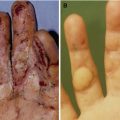
However, based on the author’s experience, allogenic dermis is difficult to be taken by the wound bed. Only a thin meshed graft can be taken. A thick non-meshed graft is more risky in terms of the wound bed accepting the allogenic dermis. In addition, resultant scar after wound healing is usually not satisfactory (Fig. 3.10).
Fig. 3.10
Thick non-meshed allogenic dermis grafts usually result in unfavorable scar contractures since they are not taken on the wound beds
The author’s technique of applying the allogenic dermis graft is as the following. Briefly, the thin meshed allogenic dermis is tailored to the size and shape of the defect and transferred to the recipient site. The transplanted allogenic dermis is fixed to the wound bed along the circumference of the defect using 6-0 Prolene sutures. Dressings are kept moist by applying fibrin glue and ointment until complete reepithelialization is achieved. The skin sutures are removed after 5–7 days. After wound healing, sunlight is avoided for the first 3–6 months using sun-blocking agents.
Artificial Dermis Graft
One of the contributing factors for unfavorable results using allogenic dermis to cover the wounds is that matrix of the allogenic dermis is too compact for the cells and tissues of the wound bed to integrate into the allogenic dermis. In order to overcome the limitation of allogenic dermis, artificial dermis can be used instead. Recently, commercially available artificial dermis has been commonly used in clinical practice. Artificial dermis should have optimal physical and biochemical factors for its successful use in regenerative medicine. Collagen and hyaluronic acid sponges/sheets are two main biomedical materials that are frequently used in clinical medicine, especially in dermatology and plastic surgery. These materials have been known to accelerate tissue granulation. Collagen and hyaluronic acid matrices promote migration of host cells and blood vessels into the structure, thus allowing rapid replacement by host tissue.
For the artificial dermis grafting, an artificial dermis is tailored to the size and shape of the defect and transferred to the recipient site in 2–4 layers. The transplanted artificial dermis is fixed to the wound bed using fibrin blue. Dressings are kept moist by applying fibrin glue and ointment until complete reepithelialization is achieved. Sunlight is avoided for the first 3–6 months using sun-blocking agents.
Related posts:
Stay updated, free articles. Join our Telegram channel

Full access? Get Clinical Tree


