(1)
Professor of Plastic Surgery, Director of Diabetic Wound Center, Director of Cell Therapy Laboratory, Korea University College of Medicine and Korea University Guro Hospital, Seoul, Republic of Korea (South Korea)
Abstract
Autologous tissues including dermis and fat have long been used for soft tissue augmentation to correct facial wrinkles or skin contour defects. Additionally, given the advances in molecular biology and tissue engineering, soft tissue filler products are widely used. However, their variable degrees of resorption require repeated injections. To overcome these drawbacks, the author created a new injectable filler consisting of a mixture of hyaluronic acid filler and living autologous cells. In this chapter, the author’s basic research and clinical experiences with the tissue-engineered injectable soft tissue using cultured dermal fibroblasts, adipose-derived stromal vascular fraction cells and adipose-derived in vitro-differentiated adipocytes are presented. The injection of tissue-engineered soft tissue has many advantages. It is a simple procedure and can be carried out at an office without entering the operation room. Moreover, it almost eliminates communication errors between the physician and patient, because patients can immediately assess the results. In addition, patients feel more comfortable because autologous tissue is used.
Keywords
Tissue engineeringSoft tissueFibroblastSVF cellAdipocyteDermis, fat, and dermis fat grafts have long been used for soft tissue augmentation to correct facial wrinkles or skin contour defects (Figs. 12.1 and 12.2). As increasing number of patients seek aesthetic improvement through minimally invasive procedures, the demand for effective and durable soft tissue fillers to correct facial wrinkles or to augment soft tissues has grown dramatically. To meet these demands, various commercially available biologic implants have been developed and are being widely used. In particular, allogenic dermis and injectable filler substances are replacing the conventional autologous tissue transfer.

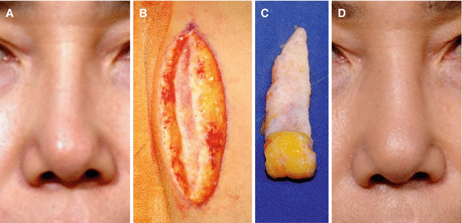
Fig. 12.1
Correction of soft tissue atrophy of the cheek. (A) Preoperative view. (B) Postoperative view after fat injection
Fig. 12.2
C-shaped nasal deviation occurred after augmentation rhinoplasty with a silicone implant. The implant was removed, and the nasal dorsum was augmented again by dermis fat grafting. (A) Preoperative view. (B, C) Harvested dermis fat. (D) Six years after the graft
As mentioned in Chap. 3, allogenic dermis has been a widely accepted acellular human dermal matrix for skin and soft tissue applications. Donated human skin is aseptically processed to remove the epidermis and cells that can lead to tissue rejection and graft failure. The implanted allogenic dermis is transformed into the host tissue, ultimately producing the effect of soft tissue augmentation by revascularization and cell migration from the host tissue. Allogenic dermis has been commonly used as a biologic implant to correct soft tissue defects or wrinkles (Figs. 12.3, 12.4, and 12.5).
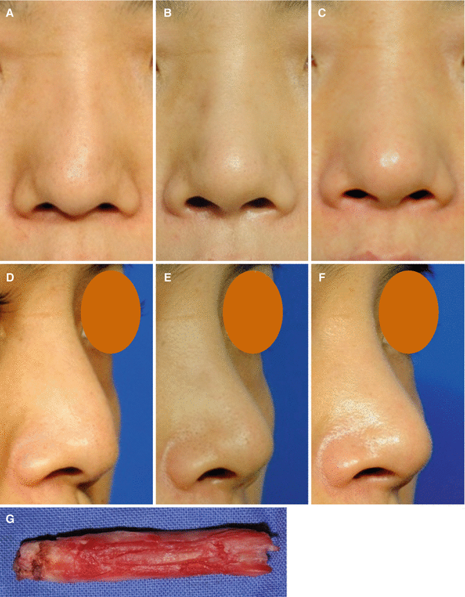
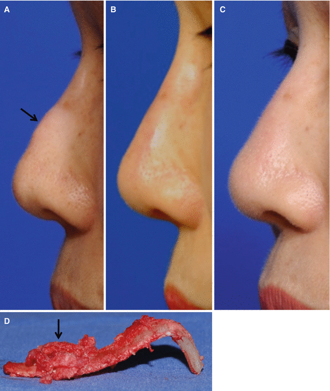
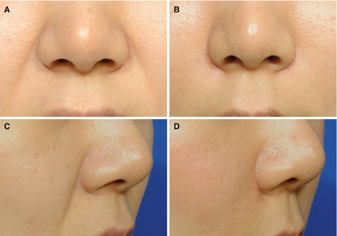
Fig. 12.3
(A) Reverse C-shaped nasal deviation occurred after augmentation rhinoplasty with a polytetrafluoroethylene (PTFE) implant. (B) The implanted PTFE was removed, and the nasal dorsum was augmented again by allogenic dermis grafting. Three months after the graft. (C) One year after the graft. (D–F) Three-quarter views. (G) The removed PTFE
Fig. 12.4
(A) A hard subcutaneous mass (an arrow) caused by calcification of an implanted silicone. (B) The calcified silicone implant was removed, and the nasal dorsum was augmented again by an allogenic dermis graft. One month after the graft. (C) One year after the graft. (D) The removed calcified (an arrow) implant
Fig. 12.5
The deep nasolabial folds were corrected by allogenic dermis grafting. (A) Preoperative view. (B) Six months after the graft. (C, D) Three-quarter views
An ideal injectable filler should be autologous and easily applied, provide long-term results, require minimal surgery, and show minimal donor site morbidity. Given the advances in molecular biology and tissue engineering, the list of natural injectable fillers continues to grow. Human (both autologous and homologous), animal (bovine and porcine), and even bacterial sources are now available. Each product has its own advantages and disadvantages. In particular, rapid resorption and disappointing overall long-term results are common problems for all available natural fillers.
Currently, soft tissue filler products based on hyaluronic acid (HA) are widely used, since they have a low potential for allergic reactions, require no skin testing, can be stored at room temperature, and have no risk of bovine spongiform encephalopathy unlike collagen (Fig. 12.6). Although HA fillers have shown to be relatively safe and convenient to administrate, their variable degrees of resorption require repeated injections. To overcome these drawbacks, the author created a new injectable filler consisting of a mixture of HA filler and living cultured human fibroblasts.
Fig. 12.6
A sunken eyelid was corrected by injection of an HA filler (arrows). (A) Preoperative view. (B) Immediate after the injection. (C) Three months after the injection
In this chapter, the author’s experiences with the tissue-engineered injectable soft tissue using autologous cells are presented.
Injectable Soft Tissue Using Fibroblasts
Autologous cultured fibroblasts have been successfully utilized as a living, dynamic protein repair system for dermal and subcutaneous deficiencies since 1995. The introduction of this technique opened a new vista of opportunities for plastic surgeons. Autologous cultured and expanded living fibroblasts create a protein repair system that appears to show continuing correction for many months to years.
Cultured human fibroblasts are added to HA fillers based on the hypothesis that the injection of extracellular matrix (ECM)-producing cells results in a longer correction effect than that achieved using an HA filler alone. The HA filler is expected to provide a dramatic early fill and allow cultured human fibroblasts to provide a long-lasting effect which is not achievable with the filler alone.
Basic Research
To develop the soft tissue augmentation method using injectable tissue-engineered soft tissue consisting of a mixture of HA filler and cultured human fibroblasts, the author performed three basic researches.
Feasibility of Injectable Tissue-Engineered Soft Tissue Using HA Filler
Natural HA provides a biological material with high viscoelastic and rheological properties which, in addition to its non-immunogenicity, biocompatibility, and total biodegradability, make it suitable for various medical applications. Thus, it was hypothesized that HA fillers could be used as injectable cell carriers and as suitable scaffolds as well as filling materials of the skin and soft tissue.
A study was undertaken to evaluate the feasibility of Restylane®, which is a modified HA, combined with cultured human dermal fibroblasts, to enhance the longevity of injected implants. The histological changes of the injected implants were also evaluated.
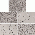
For the test group, fibroblasts from the dermis of healthy adults were isolated and cultured. Fibroblasts suspended in Dulbecco’s phosphate-buffered saline without Mg2+ or Ca2+ (DPBS) were then dispersed in Restylane® to form a human fibroblast Restylane® mix. For the control group, DPBS without fibroblasts were mixed with Restylane®. These implants were subcutaneously injected into the back of an athymic nude mouse. The nodular swellings that resulted from the injections were excised to include the skin beyond the swelling points down to the panniculus carnosus layer. The weights were measured at 1, 2, 4, 8, 12, and 16 weeks after the injections. Histological comparisons were performed to confirm the presence of human collagen in the fibroblast-mixed Restylane® group through an immunohistochemical study with antihuman collagen type I polyclonal antibody.
The mean weight of the control group nodules decreased throughout the examination period. The mean weight at the 16th week was 60 % of the weight measured at the first week. On the other hand, the mean weight of the test group nodules decreased only over the first 2 weeks. Beyond 2 weeks, there was no further significant weight change. The mean weight at the 16th week was 91 % of the weight measured at the first week. Histological examinations of the control group exhibited negative immunohistochemical staining for human collagen at each examination period. The test group exhibited positive staining after 2 weeks, indicating the presence of human collagen. Restylane® mixed with cultured human dermal fibroblasts did not produce local tissue reactions from the recipient.
These results indicate that Restylane® mixed with cultured human dermal fibroblasts may be successfully utilized as living grafts for long-term protein repair systems (Fig. 12.7).
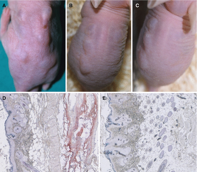
Fig. 12.7
(A–C) The implants were subcutaneously injected into the back of an athymic nude mouse at six sites, the three left sites composing the test group of fibroblasts suspended in Restylane and the three right sites composing the control group of Restylane only (A immediate after injection, B 8th weeks, C 16th weeks). (D, E) Immunohistochemistry taken at the 16th week after injection (D test group, E control group). The test group shows strong immune reactions with anticollagen type I and confirms the presence of human type I collagen (red staining)
Tracking and Increasing Viability of Fibroblasts Suspended in HA Filler After Topical Injection
To establish the injectable tissue-engineered soft tissue method as a standard treatment, a study is required to determine whether the injected fibroblasts could stay at the injected place or move to other sites. In addition, effective strategies are needed to increase viability of the injected fibroblasts.
A study was carried out to track the injected fibroblasts and to determine the effect of adding prostaglandin E1 (PGE1) or vitamin C on the viability of fibroblasts. PGE1 and vitamin C were tested since they are cheap and have been commonly used in clinical settings. PGE1 is known to mediate vasodilation and improve microcirculation perfusion. Recently, PGE1 has been also reported to stimulate fibroblast proliferation. Vitamin C is well known for promoting fibroblast production. Therefore, it was hypothesized that a combination of PGE1 or vitamin C in the fibroblast HA filler might accelerate fibroblast viability and/or proliferation after injection into the body.
Human fibroblasts labeled with fluorescence dye were suspended in HA filler and injected into four sites on the back of nude mice. The injected bioimplants consisted of one of four of the following: HA filler without cells (HA group), fibroblasts suspended in HA filler (HA + FB group), PGE1-supplemented fibroblasts in HA filler (HA + FB + PGE1 group), and vitamin C-supplemented fibroblasts in HA filler (HA + FB + VC group). At 4 weeks after injection, locations and intensities of the fluorescence signals were evaluated using a live imaging system.
The results demonstrated that the fluorescence signals of the fibroblast-containing groups were visible only at the injected sites without dispersing to other sites. The HA + FB + PGE1 group showed a significantly higher fluorescence signal than the HA + FB and the HA + FB + VC groups. There was no statistical difference between the HA + FB and HA + FB + VC groups.
The results of the study collectively suggest that injected fibroblasts suspended in HA filler stay at the injected place without moving to other sites. In addition, PGE1 treatment may increase the viability and/or proliferation of the injected fibroblasts (Figs. 12.8 and 12.9).
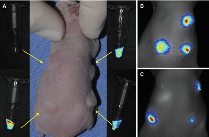
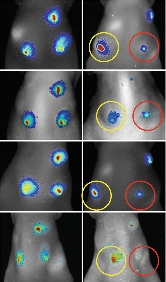
Fig. 12.8
(A) Nanoparticle-positive (labeled) human dermal fibroblasts suspended in the HA filler were injected intradermally into the back of mice at four sites, the left upper site for the group of HA filler without cells, the right upper site for the group of fibroblasts suspended in the HA filler, the left lower site for the group of PGE1-supplemented fibroblasts suspended in the HA filler, and the right lower site for the group of vitamin C-supplemented fibroblasts suspended in the HA filler (arrows). (B) An optical image obtained immediate after the injections of the bioimplants. (C) An optical image at 4 weeks after the injections. The injected fibroblasts in HA filler stayed at the injected place without dispersing to other sites
Fig. 12.9
Fluorescence signals of the labeled human fibroblasts immediately (left photos) and 4 weeks (right photos) after injection of the bioimplants in the matched cases. PGE1 supplementation (yellow circles) shows the best result in viability of the injected fibroblasts. However, vitamin C supplementation (red circles) does not significantly increase viability of the cells
Optimal Condition of HA Filler as a Carrier for Fibroblasts
Although HA fillers have been basically developed as materials used independently without being mixed with cells, recent reports have demonstrated that HAs can also be used as injectable cell carriers and as suitable scaffolds. According to the author’s studies described above, cultured human dermal fibroblasts mixed into nonanimal stabilized HA fillers can survive and produce human dermal matrices and be used as a long-lasting injectable soft tissue for augmentation to sustain the corrective effect. However, there have been few experimental reports demonstrating appropriate conditions of HA fillers as cell carriers.
The author performed a study to determine the optimal characteristics of HA filler, combined with cultured human dermal fibroblasts, to enhance the maximal viability of injected cells. The results demonstrate that the HA-based filler with moderate viscosity (2,000,000–4,000,000 centipoises) is superior to those with low (600,000–800,000 centipoises) or high (8,000,000–12,000,000 centipoises) viscosity in the viability for human fibroblasts. The shape of the particles (round or irregular) does not have an influence on the viability of the injected fibroblasts (Figs. 12.10 and 12.11).
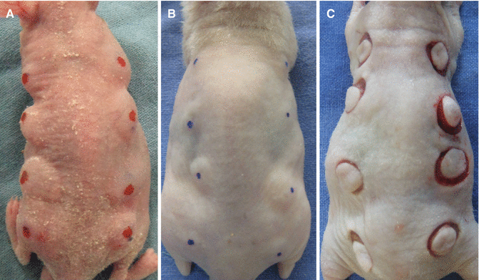
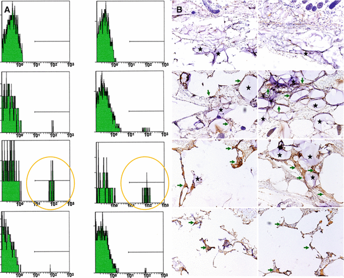
Fig. 12.10
An HA filler mixed with cultured fibroblasts was injected intradermally into the back of athymic nude mice at eight sites. The most cephalic sites composed the control group. In experimental groups, the second row was for the low viscosity group, the third row was for the moderate viscosity group, and the bottom row was for the high viscosity group. (A) Immediate after the injections. (B) At 16th week after the injections, the nodular swelling of the moderate viscosity group is most prominent. (C) Biopsy samples for FACS and immunohistochemistry
Fig. 12.11
(A) The result of FACS for human type I collagen. The photographs show that the moderate viscosity group has the greatest amount of human collagen (yellow circles). (B)The photographs of immunohistochemistry (×100). The green arrows (dark brown staining) indicate human type I collagen. The black stars are the remnant HAs. The moderate viscosity group demonstrates the greatest amount of human collagen
Application Method
Autologous dermal fibroblasts are isolated from dermal tissue harvested from a patient’s inguinal or gluteal area and are then cultured. In order to obtain a sufficient amount of fibroblasts for injection, 4–6 weeks are required.
Ten to twenty million cultured fibroblasts are suspended in 1.0–1.5 ml of an HA filler. The desired shape and size to be augmented is decided by the patient. After preparing the skin around the injection site with common antiseptic solutions, the fibroblast filler mixture is injected into the intradermal, subdermal, and/or subcutaneous layer using 23- and/or 26-gauge needle (Fig. 12.12).
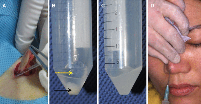
Fig. 12.12
Application methods. (A) Skin harvesting. (B) An HA filler (a yellow arrow) and a cultured fibroblast pellet (a black arrow). (C) The cultured fibroblasts are mixed with the HA filler. (D) The fibroblasts suspended in the HA filler are injected into the nasal dorsum
Immediate molding is performed with finger pressure in order to prevent possible uneven beading of the injected implant. At a point where the patient is most satisfied with the augmentation amount, an additional 0.1–0.5 ml of the bioimplant is injected to achieve an overcorrection of 30 %. The usual amount of injected implant per patient is 0.6–1.5 ml.
The implant is not injected into the nasal tip area due to the elevated densities of sebaceous glands and large skin pores.
Clinical Experience
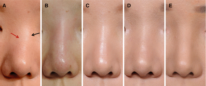
The author has applied the injectable tissue-engineered soft tissue of HA mixed with autologous-cultured fibroblasts for augmentation rhinoplasty and wrinkle correction since 2002.
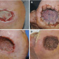
Patients show mild erythema at the injection site immediately after the procedure, but this resolves completely within 1–2 days. Patients feel that the volume of the injected implant appears to diminish during the early postoperative 2–4-month period. However, the augmentation effect is well maintained after this initial period. The amount of volume reduction is usually 20–40 %. The majority of patients are well satisfied with the natural shape and feel of augmentation (Figs. 12.13, 12.14, 12.15, 12.16, and 12.17).
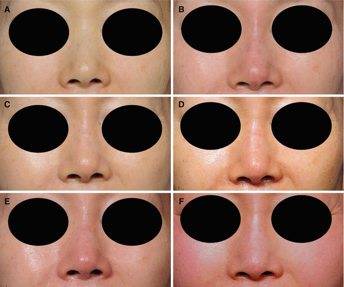
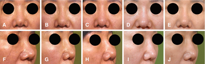
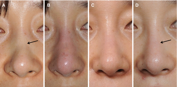
Fig. 12.13
A 59-year-old woman was treated by tissue-engineered soft tissue injection for augmentation rhinoplasty. (A) Preoperative view. (B) Immediate postoperative view. (C) Two weeks after the injection. (D–F) Six, twelve, and eighteen months after the injection
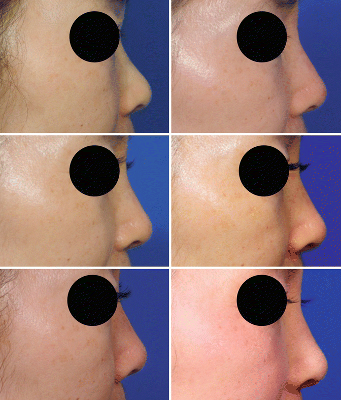
Fig. 12.15
A 49-year-old woman had previously undergone an operation for silicone implant removal due to implant extrusion. After 3 months, she was treated by tissue-engineered soft tissue injection into the nasal dorsum and for a depressed scar area at the infratip–columellar junction caused by silicone implant extrusion. (A) Preoperative view. (B–E) Two weeks, 3, 6, and 15 months after the injection. (F–J) Profile views
Fig. 12.16
A 43-year-old patient had her nasal bone broken. (A) One year after the fracture, the rhinion area was depressed (an arrow). The depression was corrected by tissue-engineered soft tissue injection. (B) Immediate after injection. (C) Six months after the injection. (D) Two years after the injection. The augmentation effect was well maintained (an arrow)