Hidradenitis suppurativa (HS) is a chronic relapsing disease of follicular occlusion that causes immense clinical and psychosocial morbidity when refractory to treatment. HS is no longer considered a disease of primary infectious etiology, although bacteria play a role. There is increasing evidence that HS is associated with immune dysregulation, based on its clinical association with other immune-mediated disorders, by its response to biologic therapy in the clinical arena, and from molecular research. This article summarizes what is known in relation to the inflammatory pathways in HS.
Key points
- •
Cytokines involved in hidradenitis suppurativa (HS) pathogenesis include interleukin (IL)-1β, IL-17, IL-10 and to a lesser extent TNF-α, therefore representing potential therapeutic targets.
- •
An overzealous toll-like receptor response to commensal bacteria may be an initiating factor in the the pathogenesis of this disease. In advanced stages, characterised by sinus tract formation and scarring, superinfection with bacterial strains known to induce soft tissue infection may occur, which frequently responds to intensive targeted antibiotic therapy.
- •
Smoking is epidemiologically linked to HS. The mechanism of action of smoking in initiating or propagating HS is theoretically by:
- ○
Inducing epidermal hyperplasia and keratinization, leading to occlusion of the follicular infundibulum
- ○
Alteration of the skin immune response
- ○
Increasing microbial virulence and decreasing skin antimicrobial peptides
- ○
Introduction
Hidradenitis suppurativa (HS) is a chronic, debilitating disease with significant associated psychosocial morbidity. Reflecting the lack of an established pathogenic pathway, treatments for HS remain suboptimal. A growing body of research and evidence, however, is paving the way for better management of patients with this difficult condition. The key to unlocking therapeutic options may be the elucidation of the inflammatory mechanisms at the core of the disease process.
Follicular occlusion, as opposed to apocrinitis, is central to the development of HS. The cause of this is likely multifactorial. Environmental triggers (eg, cigarette smoking and adiposity) in a genetically susceptible individual result in the HS phenotype. The role of aberrant immunity in HS has become topical and is suggested by an association with other immune-mediated diseases such as Crohn’s disease and pyoderma gangrenosum, and by a favorable response to tumor necrosis factor alpha (TNF-α) blockade. It is suggested that immune mechanisms contribute to follicular occlusion. This is hypothesized as innate and adaptive immune cells (and their products) are found in abundance in lesional and perilesional skin, and are thought to precede clinically apparent HS. Despite scientific advancement, the exact pathogenic pathways are poorly defined, and the precise cytokine profile in HS has not been fully elucidated.
Cytokines in hidradenitis suppurativa
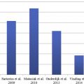
There is a lack of consensus as to which cytokines and immune pathways drive the inflammation in HS. TNF-α is a pivotal proinflammatory cytokine, produced by innate and adaptive immune cells, and it has an established role in many inflammatory conditions such as psoriasis, Crohn disease, rheumatoid arthritis, sarcoidosis, and uveitis. Because clinical improvement in HS is frequently observed with infliximab and adalimumab treatment, it is hypothesized that TNF-α expression is enhanced in this disease. Several studies have demonstrated the presence of TNF-α at mRNA and protein levels in HS skin samples compared to healthy controls, and in some cases, comparable to levels in psoriatic skin. Dréno and colleagues found that the protein concentration of TNF-α was in fact decreased in lesional and perilesional HS skin compared with healthy controls, suggesting deficient cutaneous innate immunity. A limitation of this study however, is that conclusions were drawn based on immunohistochemical analysis using paraffin-embedded tissue sections only, which limit firm interpretation. Other studies looked at circulating concentrations of TNF-α in peripheral blood/serum, again with conflicting results. Giamarellos-Bourboulis and colleagues demonstrated a diminished innate response to lipopolysaccharide stimulation in HS, with monocytes from patients producing less TNF-α than healthy controls. A possible explanation of these in vitro results may be that they are caused by in vivo anti-inflammatory priming of HS monocytes (eg, by the high interleukin [IL]-10 levels in HS). In contrast, Matusiak and colleagues demonstrated enhanced concentrations of TNF-α in HS serum. In a study by van der Zee and colleagues looking at the effects of adalimumab on cytokine expression in HS skin, biopsies were taken before and at week 16 after treatment. The protein expression of TNF-α and its receptors was enhanced in HS skin before treatment, but not as prominently as other cytokines such as IL-1β and IL-10 when compared with healthy control skin (1.6-fold increase vs 54 and 14.8 respectively). Similarly, the response to adalimumab was not as marked on TNF-α concentrations than these other cytokines. Another recent study demonstrated enhanced mRNA expression of TNF-α in lesional and perilesional HS skin, but less pronounced that the enhancement of IL-17 and IL-1β. This suggests that TNF-α, although present in inflammatory HS skin, may not be one of the big players in the pathogenesis of this disease. Overall, there is conflicting evidence in relation to the role of TNF-α in HS.
The data for IL-1β in HS, albeit from fewer studies, seem to be more consistent with IL-1β often one of the most enhanced cytokines demonstrated in various studies. IL-1β is a potent proinflammatory cytokine that drives the differentiation of Th17 cells. Mature IL-1β is synthesized from its inactive precursor following cleavage by activated caspase-1, which in turn requires the assembly of a multiprotein complex known as the inflammasome. Activation of the inflammasome is implicated in several inflammatory disorders such as gout. A 31-fold enhancement of IL-1β was demonstrated in HS lesional skin, and elevated concentrations were also observed in perilesional skin. In another study by the same authors, a 54-fold increase in protein expression of IL-1β was observed. A 115 fold enhancement of the mRNA expression of IL-1β in lesional and perilesional HS skin has recently been demonstrated and in this study it has been suggested that a subset of CD14+ dermal dendritic-cells produce IL-1β. Wolk and colleagues also documented enhanced mRNA expression of IL-1β, exceeding that of psoriatic skin. Meanwhile, the anti-inflammatory cytokine IL-10 has also been observed frequently in HS skin. Its presence in involved skin is likely a response to a proinflammatory environment, although it is conceivable that high levels in lesional skin may have local immunosuppressive effects, therefore propagating inflammation and infection. Known cellular sources of IL-10 are activated T cells, macrophages, mast cells, and B lymphocytes that are abundantly present in HS lesional skin.
The role of IL-17 in HS pathogenesis is emerging. IL-17 is produced by Th17 cells, which have a central role in many autoimmune diseases such as psoriasis and Crohn’s disease. Besides Th17 cells, innate lymphoid cells, γ-δ T cells, mast cells, and neutrophils have recently been shown to produce IL-17 also. Th17 cell development is driven by IL-1β, IL-6, transforming growth factor-β, and IL-23, cytokines produced by activated innate cells such as dendritic cells. Activated T cells, mast cells, and neutrophils are abundantly present in HS lesional skin. Schlapbach and colleagues demonstrated IL-17 producing CD4+ T helper cells in HS lesional dermis, with protein expression of IL-17 enhanced by a factor of 30 compared with healthy controls. The same group also demonstrated that IL-23 was abundantly expressed by macrophages infiltrating the dermis in lesional HS skin. Wolk and colleagues observed enhanced mRNA expression of IL-17 by a factor of 7, compared with control skin and similarly van der Zee and colleagues documented a seven-fold enhancement in the protein expression of IL-17 analyzed following transwell culture. A recent study also demonstrated a 149-fold enhancement of mRNA expression of IL-17 in lesional skin. In addition, enhancement of IL-17 was documented in perilesional and clinically uninvolved skin (10 cm away from an active lesion) in HS patients, suggesting a primary role for immune dysregulation in HS. CD4+ T cells were again documented as the cellular source of IL-17 in this study. The detection of IL-1β, IL-23, and IL-17 therefore implicates the IL-1β-IL-23/Th17/IL-17 pathway in the pathogenesis of this disease.
IL-6 is a pleotropic key proinflammatory cytokine, but its role in HS pathogenesis remains undefined. The IL-12/Th1 pathway was once considered pivotal in many inflammatory conditions until the discovery of Th17 cells. IL-12 has also been observed in a limited number of studies in HS skin. Remarkably however, interferon-γ, largely produced by Th1 cells, was not consistently seen in HS studies.
Antimicrobial peptides
Constitutively produced antimicrobial peptides (AMPs), released as part of the body’s innate immune response to microbial threat, are secreted by keratinocytes in large quantities in the presence of invasive pathogens. In addition to antimicrobial properties, they also have immunomodulatory effects such as cytokine production, antigen presentation, recruitment of immune cells, and wound healing. AMPs are particularly important where the skin barrier function is disrupted as a means of preventing or limiting cutaneous infection.
Several studies have evaluated AMPs in HS. All studies on AMPs in HS have limitations, because in all cases mRNA and protein expression were not measured in parallel in the same sample using robust techniques such as quantitative polymerase chain reaction (PCR) and enzyme-linked immunosorbent assay (ELISA). Wolk and colleagues demonstrated a relative deficiency, at an mRNA level, of all measured AMPs (beta defensin [BD] -1, BD-2, BD-3, S100A7 [psoriasin], S100A8 [calgranulin A], and S100A9 [calgranulin B]) in HS lesions, and this attenuation was even broader than that seen in atopic dermatitis (AD), a condition long associated with suppressed AMP levels, hence the propensity for bacterial superinfection in AD. These authors relate this to a relative deficiency of IL-22 and IL-20 in HS. Schlapbach and colleagues reported a relative deficiency of BD-2, thus facilitating colonization by bacteria. Enhanced mRNA expression of psoriasin was observed by this group, however. Hofmann and colleagues also demonstrated suppressed BD-3 and RNase-7. Relative deficiencies of AMPs, namely BD-4 and BD-2, were also documented by other groups in lesional skin compared with healthy controls. In contrast to this, however, Emelianov and colleagues demonstrated enhancement of LL-37 (cathelicidin), psoriasin, and BD-3. Overall, the pattern of AMP aberrancy is unclear in HS, but there appear to be alterations in levels and functioning of AMPs. How they contribute to the initiation/propagation of HS remains to be elucidated.
Proposed involved signaling pathways in hidradenitis suppurativa
Pathogen recognition receptors (PRRs) are germline-encoded, nonclonal binding sites for the recognition of microbes/infectious agents by the innate immune system. PRRs may be found in the cytoplasm or cell surface of immunocytes, or be secreted into the local and circulating fluids. PRRs include toll-like receptors (TLRs), the nucleotide oligomerization domain (NOD) proteins, the RIG-like helicases, and the C type lectins. The main functions of PRRs include proinflammatory signaling, complement activation, opsonization, phagocytosis, and regulation of apoptosis. They recognize microbial motifs or pathogen-associated molecular patterns (PAMPs), including bacterial wall components, viral nucleic components, various proteins of flagellae, and fungal cell wall components.
Several groups have investigated the role of TLRs in HS with varying results. Hunger and colleagues reported enhanced expression of TLR2 at mRNA and protein levels, as well as C-type lectin in the epidermis and dermis. In contrast to this, Dréno and colleagues suggested deficient innate immunity with suppressed expression of TLR2, TLR4, TLR3, TLR7, and TLR9 in HS skin compared to healthy controls. TLR expression in HS is therefore not fully defined. The role of bacteria in the initiation or propagation of HS is also unclear. Despite the clinical appearance of infection, bacterial examination, commonly from superficial lesions, has frequently shown a mixed growth of commensal microbes. This has been challenged, however, by a recent report whereby specific pathogens were isolated after broad and prolonged culturing. Specific antibiotic therapy resolved these cases of HS. In line with this, a recent case report of long-standing refractory HS demonstrated an excellent clinical response to intensive intravenous linezolid and meropenem, yet disease flared quickly after withdrawal, implying that infection is not the primary etiology of this disease. The suggestion is that there may be an overzealous TLR response to commensal bacteria as an initiating factor in the pathogenesis of HS, launching an inflammatory cascade. Once HS is established, with sinus tract and fistula formation, secondary bacterial superinfection is likely to play a role in disease exacerbation, where bacterial colonies are harbored in these tracts in a biofilm formation, making eradication challenging and propagating inflammation. Recent reports suggest that, at least in advanced stages of HS, superinfection with bacterial strains known to induce soft tissue infections may occur, which responds to intensive targeted antibiotic therapy.
Advances in the genetics of HS have demonstrated loss of function mutations in 3 of the 4 subunits of γ-secretase, at least in a clinical subtype of familial HS. γ-secretase cleaves the intracellular domain of Notch and Notch- signaling has many functions including the regulation of hair follicle differentiation and maintenance of the epidermal barrier. Defective Notch signaling therefore occurs in HS, resulting in the inhibition of the hair growth cycle, the conversion of hair follicles into keratin-enriched epidermal cysts, and poor sebaceous gland differentiation. Notch and TLR interaction has been reported, in which notch signaling suppresses TLR-4 induced proinflammatory cytokine responses by macrophages. As this negative feedback is impaired with Notch deficiency a proinflammatory environment ensues. IL-22 deficiency, demonstrated previously in HS may be linked to Notch deficiency, as IL-22 secretion by T cells is Notch dependent ( Box 1 ).
Related posts:
 Prevalence, Risk Factors, and Comorbidities of Hidradenitis Suppurativa
Diagnosing Hidradenitis Suppurativa
Prevalence, Risk Factors, and Comorbidities of Hidradenitis Suppurativa
Diagnosing Hidradenitis Suppurativa
 The Handicap of Hidradenitis Suppurativa
Endocrinologic Aspects of Hidradenitis Suppurativa
The Handicap of Hidradenitis Suppurativa
Endocrinologic Aspects of Hidradenitis Suppurativa
 Randomized Controlled Trials for the Treatment of Hidradenitis Suppurativa
Imaging of Hidradenitis Suppurativa
Randomized Controlled Trials for the Treatment of Hidradenitis Suppurativa
Imaging of Hidradenitis Suppurativa
Stay updated, free articles. Join our Telegram channel

Full access? Get Clinical Tree


