Hidradenitis suppurativa is a complex disease of chronic evolution and difficult management. Imaging, particularly color Doppler ultrasound, has demonstrated a wide range of subclinical anatomic abnormalities, allowing modification of the clinical assessment of severity of the disease and therefore management of patients. Sonography supports early and more precise diagnosis and staging by providing critical objective information in real time. The richness of these data can also support assessment of the pathogenesis of the disease, allow monitoring of patients, and contribute to clinical trials. MRI can support the diagnosis of extensive anogenital and deep lesions.
Key points
- •
Clinical examination alone can underestimate the severity of HS.
- •
Color Doppler ultrasound may support the management of hidradenitis suppurativa.
- •
Sonographic criteria for supporting the diagnosis are established.
- •
Disease staging is possible using ultrasound.
- •
MRI can help in the diagnosis of extensive and deep anogenital lesions.
Introduction
Hidradenitis suppurativa (HS) is a complex disease of difficult management. The actual origin of the disease remains unclear. Moreover, according to a review by Rambhatla and colleagues most therapies used to treat HS are only supported by limited or weak scientific evidence. Even though the diagnosis is mainly based on visual inspection, clinical assessment of the severity presents limitations. Thus, the usually used Hurley scoring relies only on clinical data, but lacks the accurate anatomic information necessary for use in clinical trials. The Sartorius scoring seems to be time-consuming and relies on some clinical data that may be inaccurate and need modification after the use of imaging, such as the longest distance between two lesions separated by clinically normal skin. Clinical interpretation of the nodules, abscesses, and fistulae may also be subject to reinterpretation under imaging. Palpation seems to be limited spatially because of the strong presence of inflammation and it may lead to misinterpretation of a fistula for a nodule, which can produce an erroneous assignment of the stage of severity and therefore the treatment. Histology seems to be limited in HS because of the extensive and deep multiregional involvement.
The role of imaging in HS is to detect the actual type and extent of anatomic abnormalities by providing objective and precise information. Hence, it should support early diagnosis and assessment of severity of the disease. This may also include monitoring of treatment, presurgical mapping of lesions, and provision of detailed anatomic information for clinical trials. Furthermore, it has been demonstrated that the addition of imaging, particularly color Doppler ultrasound, can significantly modify management in more than 80% of HS patients because of the clinical underestimation of severity.
The first-line and most frequent imaging examination used for studying HS is color Doppler ultrasound. This imaging technique presents good resolution for studying the skin and deeper layers. MRI has been mainly used in some extensive and deep anogenital HS lesions. Nevertheless, MRI is expensive and currently the commercially available units present limitations in their resolution for detecting abnormalities in skin layers. However, it can observe the anatomic alterations in deeper structures.
As with any medical test, the performance of imaging examinations requires adequate equipment and training of the operators that acquire the images and interpret the examinations. The latter is of paramount importance for standardizing the acquisition protocols and the categorization of lesions.
Ultrasound
This is an imaging modality based on the propagation and return of sound waves in different tissues. Ultrasound is a real-time and safe imaging method without reported adverse reactions. Usually, it does not require intravenous injection of contrast media and allows a rich and live interaction between the sonographer and patient. The structures or tissues can be classified according to their echogenicity pattern into anechoic (ie, fluid-filled structures that allow the passage of the sound waves; black), hypoechoic (ie, dense fluid-filled structures or solid structures; gray), and hyperechoic (ie, solid structures that might present capillary vascularity, heavy calcium deposits, or exogenous materials; white). Each of the echogenicity categories presents variations according to a gray scale map and the structures are also identified according to some posterior artifacts that depend on the nature of the lesion or structure. For example, fluid-filled lesions, such as cysts, present a posterior enhancement acoustic artifact (ie, increased transmission of the sound waves; a lighter area deep to the lesion). In contrast, strongly calcified lesions show a posterior acoustic shadowing artifact (ie, lack of transmission of the sound waves; a black shadow deep to the structure). On color Doppler or power Doppler (ie, for slow flow detection) examination the blood flow patterns can be described in real time, which includes the type (arterial or venous) and the velocity of the flow (spectral curve analysis in centimeter per second).
Ultrasound is a multiaxial imaging examination that can measure the structures in centimeters or millimeters along any axis. This can be useful in HS where, for example, the fistulous tracts tend to show a tortuous distribution that can follow several axes.
Nowadays, the most common types of ultrasound machines for studying HS are multichanneled equipment with high and variable frequency probes that vary in their upper frequency range between 15 and 22 MHz. The main advantages of these machines are their good balance between resolution and penetration, which allows study of the skin and deeper layers without losing definition. Additionally, their color Doppler capabilities allow clinicians to observe and follow the patterns of the lesional and perilesional vascularity, which can be signals for detecting and monitoring activity. Currently, the limitations of this equipment are the detection of epidermal-only lesions, measuring less than 0.1 mm, and the detection of pigments. Neither of these limitations seems particularly relevant for HS because of the usually dermal and hypodermal affection reported in this disease.
On ultrasound, normal hair follicles appear as hypoechoic oblique dermal bands and the hair tracts present a predominantly trilaminar hyperechoic appearance in the scalp. However, in the rest of the body they tend to show a bilaminar or monolaminar hyperechoic linear pattern, which is compatible with the villus type of hair.
The usage of ultrasound in HS started with 20-MHz compact ultrasound units that lack color Doppler during the 1990s. These allowed detection of the widening of the hair follicles in HS patients. Later, with the development of high-frequency multichanneled color Doppler ultrasound machines the detection of subclinical abnormalities, such as echostructural dermal alterations (ie, increased thickness and decreased echogenicity), and the presence of anechoic or hypoechoic dermal and hypodermal fluid collections and fistulae were added to the detection of dilatation of the dermal hair follicles.
Interestingly, on sonography it has been demonstrated that enlarged lymph nodes are rarely found in HS, and only appear involved in late and usually severe stages. It seems that this enlargement (ie, >1 cm transverse axis) of the lymph nodes is mainly secondary to an infection rather than the disease by itself.
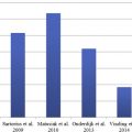
In 2010, Kelekis and colleagues showed in a small series that ultrasound can be used for supporting the diagnosis and assessing the severity of the disease through registering two parameters: dermal thickness and echogenicity.
In 2012, the usage of three-dimensional ultrasound reconstructions in HS demonstrated early and more detailed morphologic alterations in the hair follicles, such as the widening of their bases (“champagne bottle sign”), the gathering of two or more hair follicles through their basal parts, the consistent communication of the fluid collections and fistula to the widened hair follicles, and the early development of fistulous tracts that depart from the base of the hair follicles and run through the dermis and hypodermis.
In 2013, 16 years after the first report of ultrasound examination in HS, the ultrasound criteria for diagnosing HS and a sonographic scoring system for assessing severity in HS (SOS-HS) were proposed. This was mainly based on the presence of anatomic alterations, such as dermal echostructural abnormalities, and the recognition of dermal and/or hypodermal fluid collections and fistulae. Also, increased blood flow through low-velocity (≤15 cm/s) arterial and/or venous vessels was frequently detected in the periphery of these fluid collections and fistulae.
Another interesting sonographic finding is that patients’ assessments of flare activity, pain, and erythema seem to be strongly associated with morphologic changes identified using ultrasound, suggesting that these items might be strong indicators of the degree of inflammation present in HS.
As recently reported, the presence of retained fragments of hair tracts within the fluid collections and fistulae is an extremely common finding ( [CR] ; available online at http://www.derm.theclinics.com ). These variable size ectopic hair fragments seem to have lost their normal perpendicular vertical growth and outgrowth direction, and instead follow the axis of the skin layers. Probably, the presence of these nonreabsorbable keratin components that the human body does not recognize as foreign material allows the continuity and progression of the inflammatory process. Their actual role in the pathogenesis of the disease still remains to be determined; however, they seem to be linked to the persistent chronic evolution and the severity of HS.
Ultrasound Criteria for Hidradenitis Suppurativa Diagnosis
The sonographic criteria for diagnosing HS are based on the following findings: widening of the hair follicles, dermal alterations, pseudocysts, fluid collections, and fistulae. The presence of three or more findings is the sonographic criteria for diagnosing HS ( Box 1 ; Figs. 1–8 ) ( Videos 1–3 ; available online at http://www.derm.theclinics.com ).
Related posts:
 Prevalence, Risk Factors, and Comorbidities of Hidradenitis Suppurativa
Diagnosing Hidradenitis Suppurativa
Prevalence, Risk Factors, and Comorbidities of Hidradenitis Suppurativa
Diagnosing Hidradenitis Suppurativa
 The Handicap of Hidradenitis Suppurativa
Endocrinologic Aspects of Hidradenitis Suppurativa
Inflammatory Mechanisms in Hidradenitis Suppurativa
The Handicap of Hidradenitis Suppurativa
Endocrinologic Aspects of Hidradenitis Suppurativa
Inflammatory Mechanisms in Hidradenitis Suppurativa
 Randomized Controlled Trials for the Treatment of Hidradenitis Suppurativa
Stay updated, free articles. Join our Telegram channel

Full access? Get Clinical Tree


