Fig. 16.1
Tinea corporis. Erythematous plaque with elevated border
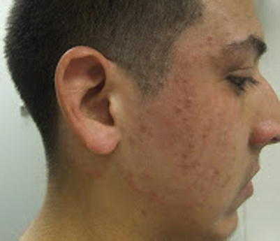
Fig. 16.2
Majocchi’s granuloma. Folliculocentric papules and pustules within a thin plaque with an expanding, elevated border on the cheek of an adolescent male
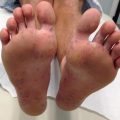
Tinea pedis is common in adolescents but is very rare in prepubertal children, and most commonly presents as interdigital scaling, erythema, maceration, or erosion. Erythema and scaling of the plantar surfaces, sometimes with vesicles and pustules (inflammatory or bullous tinea pedis) are also seen. Tinea cruris is associated with tinea pedis, and presents with pruritic thin patches or plaques with well-demarcated borders that involve the groin but typically spare the scrotum.
Tinea manuum (dermatophytosis of the palms), like onychomycosis, is rare in children, and usually presents as unilateral scaling erythema.
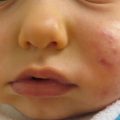
Tinea faciei involves the face, with similar clinical characteristics to that of tinea corporis (Figs. 16.3 and 16.4). Tinea barbae is a variant that involves the beard or facial hair of adolescent males.
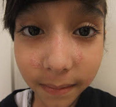
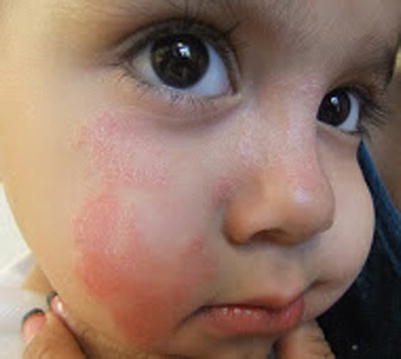
Fig. 16.3
Tinea faciei. Scaling thin plaque with elevated border on the mid-face of a young girl
Fig. 16.4
Tinea faciei. Unilateral thin erythematous plaque with central clearing in an infant
Tinea capitis is very common in the pediatric population and may be seen in up to 8 % of inner-city school-age children. The typical presentation is circumscribed patches of alopecia with scaling, erythema, and broken hairs (“black dot” of endothrix infection). Pustules may be present. Cervical or occipital lymphadenopathy is a helpful clue to the diagnosis. Kerion is a variant characterized by markedly inflamed plaque or nodule with exuberant pustules and crust (Figs. 16.5 and 16.6).
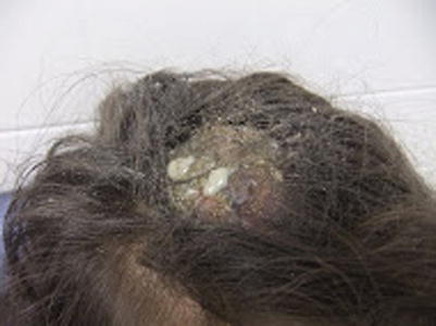
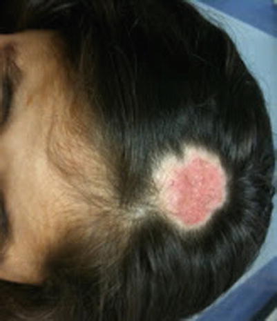
Fig. 16.5
Kerion. Large nodule with crusting and purulent drainage on the scalp of a young girl
Fig. 16.6
Kerion. Resolution of nodule and purulence following 1 month of oral terbinafine. Regrowth of hair was observed at follow-up
Onychomycosis may be due to dermatophyte, yeast such as Candida, or non-dermatophytic molds. Although rare in children, the most common cause of onychomycosis in children is dermatophytosis. Distal lateral and proximal subungual disease are characterized by yellow to white discoloration and subungual debris (Fig. 16.7). Superficial white onychomycosis is an infection of the nail plate alone.
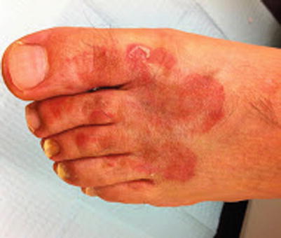
Fig. 16.7
Onychomycosis and tinea corporis. Yellow, dystrophic toenails and thin erythematous plaque on the dorsal foot
Specific Investigations
For diagnosis |
Potassium hydroxide (KOH) scraping |
Fungal culture |
Wood’s lamp (tinea capitis) |
Dermoscopy (tinea capitis) |
Histopathology |
For treatment (with systemic antifungals) |
Complete blood count (CBC) |
Liver function tests (LFTs) |
For dermatophytosis of the skin and hair, KOH preparation may be obtained by scraping active scale with a glass slide, and demonstrates a high sensitivity for diagnosis. Of note, KOH is insensitive in Majocchi’s granuloma due to the potential absence of hyphal elements in the stratum corneum. Fungal cultures allow confirmation of fungal infection within a few days and eventual speciation, but are less sensitive than KOH and take several weeks to grow on Sabouraud dextrose agar, Mycosel agar, or dermatophyte test medium. They are obtained by scraping of scale or hair with a disposable toothbrush, cytobrush, or sterile swab. Based on studies of patients with onychomycosis, KOH demonstrates a sensitivity of 76–94 % and specificity of over 70 % while culture demonstrates a sensitivity of 53–59 % and specificity of over 80 %. Histopathologic examination (nail clipping with PAS) is the gold standard for diagnosis of onychomycosis, with a sensitivity of 80–98 % and a specificity over 70 %. Skin biopsy (for histopathology, with or without PAS) is rarely necessary, but may be helpful in cases of Majocchi’s granuloma, or in cases simulating inflammatory dermatoses [3–5].
For tinea capitis, Wood’s lamp examination is helpful in cases of Microsporum infection, which demonstrates bright green fluorescence in a dark room. Given that fungal cultures take several weeks for speciation, empiric systemic antifungal therapy is recommended in clinically suspected cases of tinea capitis in children, and that terbinafine is superior for Trichophyton species while griseofulvin is superior for Microsporum species, the Wood’s lamp is a very useful adjunctive diagnostic method that allows selection of empiric therapy in tinea capitis. Dermoscopy of hair and scalp (trichoscopy) may be a useful adjunctive measure. Findings include broken hairs, dystrophic hairs, corkscrew hairs, comma hairs, black dots, horizontal white bands in hair shafts and translucent, easily deformable hairs [6–8].
In otherwise healthy children, baseline laboratory evaluation is not required prior to treatment with systemic antifungals for dermatophytosis. In children with pre-existing liver dysfunction or hematologic abnormalities, however, LFTs or CBC should be evaluated, respectively. Additionally, these tests should be performed in otherwise healthy children with lengthy courses of treatment: greater than 8 weeks of griseofulvin, 6 weeks of terbinafine, or 4 weeks of azole therapy [9].
Tinea Capitis and Barbae
Management Strategy
Treatment requires oral antifungal therapy; topical antifungals are unable to adequately penetrate follicles and should be avoided. Terbinafine and griseofulvin are the only FDA-approved medications for the treatment of tinea capitis.
Medication | Dosing | Evidence level |
|---|---|---|
Terbinafine | Granules: treatment duration for 6 weeks | A |
<25 kg: 125 mg/day | ||
25–35 kg: 187.5 mg/day | ||
>35 kg: 250 mg/day | ||
Tablets: treatment duration for 6 weeks | A | |
10–20 kg: 62.5 mg/day | ||
20–40 kg: 125 mg/day | ||
>40 kg: 250 mg/day | ||
Griseofulvin | Microsize (tablets or suspension) | A |
20–25 mg/kg/day for 6–12 weeks with maximum daily dose 1,000 mg | ||
Ultramicrosize (tablets only) | A | |
10–15 mg/kg/day for 6–12 weeks with maximum daily dose 750 mg |
Terbinafine and griseofulvin have both shown efficacy in the treatment of tinea capitis, with terbinafine showing greater efficacy for Trichophyton species and griseofulvin for Microsporum species. In one study using both agents for 6 weeks, complete cure and mycologic cure were higher for terbinafine than for griseofulvin (45.1 % vs 39.2 % and 61.5 % vs 55.5 %, respectively); however, in two meta-analyses, no significant difference in efficacy was identified. Both meta-analyses, however, did show terbinafine was more effective in the treatment of Trichophyton species while griseofulvin was more effective for Microsporum species. Additionally, terbinafine was administered for a shorter mean duration (4 weeks) than griseofulvin (8 weeks).
Medication | Dosing | Evidence level |
|---|---|---|
Fluconazole | 6 mg/kg/day × 3–6 weeks with maximum daily dose 400 mg | A |
6 mg/kg/week × 6–12 weeks (pulse regimen) | B | |
Itraconazole | 3–5 mg/kg/day × 4–6 weeks with maximum daily dose 400 mg | A |
3–5 mg/kg/day × 1 week each month for 2–3 months (pulse regimen) | B |
In one randomized-controlled trial, fluconazole for 3 weeks (6 mg/kg/day), fluconazole for 6 weeks (6 mg/kg/day), and griseofulvin for 6 weeks (11 mg/kg/day microsize formulation) all produced similar and low mycologic (44.5 %, 49.6 %, and 52.2 %, respectively) and clinical cure rates. In another randomized trial, fluconazole for 4 weeks was 78 % effective, while griseofulvin for 6 weeks was 76 % effective in curing tinea capitis. In a study comparing multiple oral antifungal agents in the treatment of T. tonsurans or T. violaceum, efficacy percentages were 92 % for griseofulvin (6 weeks), 94 % for terbinafine, 86 % for itraconazole and 84 % for fluconazole (each taken for 2 weeks). Six weeks of itraconazole 100 mg daily and ultramicrosize griseofulvin 500 mg daily produced 88 % cure rates in a patient cohort in which 90 % of the tinea capitis infections were due to M. canis.
Medication | Dosing | Evidence level |
|---|---|---|
Antifungal shampoo: | Affected patients and close or household contacts: twice weekly for 8 weeks | A |
Selenium sulfide 1 % | ||
Ciclopirox 1 % | ||
Ketoconazole 2 % | B | |
Systemic glucocorticoids (kerion) | Prednisone 0.5–1 mg/kg/day for 1 week | E |
Both selenium sulfide and ciclopirox shampoo, when used in combination with griseofulvin, demonstrated over 90 % mycologic cure rate. While one study of three children with kerion [19] demonstrated a benefit to oral steroid use, a larger retrospective study [20] found no benefit to oral or intralesional steroids.
Tinea Corporis, Faciei, and Cruris
Medication | Dosing | Evidence level |
|---|---|---|
Topical allylamines: | Once to twice daily for 2–4 weeks | A |
Terbinafine 1 % cream | ||
Naftifine 1 % cream or gel | ||
Topical benzylamine: | Once daily for 2 weeks | B |
Butenafine 1 % cream | ||
Oral terbinafine | Treatment duration: 1–2 weeks | A* |
Granules: | ||
<25 kg: 125 mg/day | ||
25–35 kg: 187.5 mg/day | ||
>35 kg: 250 mg/day | ||
Tablets: | ||
10–20 kg: 62.5 mg/day | ||
20–40 kg: 125 mg/day | ||
>40 kg: 250 mg/day | ||
Itraconazole | Treatment duration: 1 week | A |
3–5 mg/kg/day with maximum daily dose 200 mg |
In a large, pooled, meta-analysis, naftifine and terbinafine were the most effective agents in achieving mycologic and clinical cures, although efficacy was similar between azoles and allylamines. For tinea cruris, following 2 weeks of topical treatment, butenafine was over 79 % effective for clinical cure while terbinafine was 62 % effective. Mycologic cure rates were superior for butenafine compared to terbinafine (94 % and 62 %, respectively) as well. Additionally, treatment response was more rapid with butenafine. In a study of systemic treatment of tinea corporis, terbinafine was superior to griseofulvin (87 versus 73 % effective). Another study of tinea corporis comparing itraconazole to griseofulvin showed a 91 % response rate to itraconazole versus 64 % response rate to griseofulvin. Itraconazole was also significantly more useful in obtaining mycologic cure (87 % versus 57 %).
Medication | Dosing | Evidence level |
|---|---|---|
Topical azoles: | Duration: 2–4 weeks | A |
Clotrimazole 1 % cream | Twice daily | |
Econazole 1 % cream | Once daily | |
Ketoconazole 2 % cream | Once daily | |
Oxiconazole 1 % cream | Once or twice daily | |
Sertaconazole 2 % cream | Twice daily | |
Ciclopirox 0.77 % cream | Twice daily | A |
Tolnaftate 1 % cream | Twice daily | A |
Fluconazole | Duration: 2–4 weeks | A* |
6 mg/kg once weekly | ||
Griseofulvin | Duration: 2–4 weeks | A |
Microsize formulation 10–20 mg/kg/day | ||
Ultramicrosize formulation 5–15 mg/kg/day |
Tinea Pedis and Manuum
Medication | Dosing | Evidence level |
|---|---|---|
Topical allylamines: | Once to twice daily × 4 weeks | A |
Terbinafine 1 % cream | ||
Naftifine 1 % cream or gel | ||
Topical benzylamine: | Twice daily for 4 weeks | A |
Butenafine 1 % cream | ||
Oral terbinafine | Treatment duration: 2 weeks | A |
Granules: | ||
<25 kg: 125 mg/day | ||
25–35 kg: 187.5 mg/day | ||
>35 kg: 250 mg/day | ||
Tablets: | ||
10–20 kg: 62.5 mg/day | ||
20–40 kg: 125 mg/day | ||
>40 kg: 250 mg/day | ||
Itraconazole | Treatment duration: 1 week | A |
3–5 mg/kg/day with maximum daily dose 200 mg |
Allylamines and butenafine were the most effective topical agents for curing tinea pedis. However, allylamines were only slightly more effective than azoles. Ciclopirox, tolnaftate, and undecanoates were less effective than azoles and allylamines.
A systematic review demonstrated allylamines to be slightly more effective than azoles in curing tinea pedis, and azoles were more effective than tolnaftate.
Itraconazole and oral terbinafine were equally effective in curing tinea pedis. Terbinafine was significantly more effective than griseofulvin, while no significant difference was found between fluconazole and itraconazole.
Medication | Dosing | Evidence level |
|---|---|---|
Topical azoles: | Duration: 4 weeks | A |
Clotrimazole 1 % cream | Twice daily | |
Econazole 1 % cream | Once daily | |
Ketoconazole 2 % cream | Once daily | |
Oxiconazole 1 % cream | Once or twice daily | |
Sertaconazole 2 % cream | Twice daily | |
Ciclopirox 0.77 % cream | Twice daily | A |
Tolnaftate 1 % cream | Twice daily | A |
Fluconazole | Duration: 2–6 weeks | A |
6 mg/kg once weekly | ||
Griseofulvin | Duration: 4–8 weeks | A |
Microsize formulation | ||
10–20 mg/kg/day | ||
Ultramicrosize formulation | ||
5–15 mg/kg/day |
Dermatophytic Onychomycosis (Tinea Unguium)
Management Strategy
While onychomycosis is common in adults, it is less common in children under the age of 18 years. Thus, the evidence available from high-quality trials involves adult patients. Nonetheless, mycologic cure rate percentages (in descending order of superiority), based on standard dosing regimens for adults, are 76 % for terbinafine, 63 % for itraconazole pulse therapy, 60 % for griseofulvin, 59 % for itraconazole continuous therapy, and 48 % for fluconazole [31].
Medication | Dosing | Evidence level |
|---|---|---|
Terbinafine | Toenails: 12 weeks | C/A* |
Fingernails: 6 weeks | ||
10–20 kg: 62.5 mg/day | ||
20–40 kg: 125 mg/day | ||
>40 kg: 250 mg/day | ||
Itraconazole | Toenails: 12–16 weeks | C/A* |
Fingernails: 18–26 weeks | ||
<20 kg: 5 mg/kg/day | ||
20–40 kg: 100 mg daily | ||
40–50 kg: 200 mg daily | ||
More than 50 kg: 200 mg twice daily | ||
Fluconazole | Toenails: 18–26 weeks | C/A* |
Fingernails: 12–16 weeks | ||
3–6 mg/kg per weekly dose |
A prospective review of onychomycosis in children demonstrated the rarity of this condition under 18 years of age. Within a small sample size of 17 patients, oral agents were superior to topical agents. Among the oral agents, terbinafine was superior to itraconazole, which was superior to fluconazole. Of note, the evidence level for the use of these systemic agents in adults with onychomycosis is A.
In a study of children under 18 years older using ciclopirox 8 % nail lacquer solution, 77 % achieved mycologic cure and 71 % had clinical response. In adults, efinaconazole 10 % solution demonstrated a mycologic cure rate up to 55.2 % and complete cure up to 17.8 % following 48 weeks of daily treatment in multicenter, randomized, double-blind studies.
In an open trial of an over-the-counter mentholated ointment, Vicks Vaporub (eucalyptus oil, camphor, menthol, thymol, and oils of turpentine, nutmeg and cedar leaf), 27.8 % of patients had a clinical and mycologic cure following 48 weeks of daily application. The Nd:Yag 1,064-nm laser, delivered over four or eight weekly treatments, was up to 68 % effective in terms of clinical response. Both studies were performed in adult patients only.
Tinea Versicolor
Clinical Features
Tinea versicolor or pityriasis versicolor is caused by organisms of Malassezia genus, which are normally commensal, but become pathologic when the yeast form transforms into mycelia under conditions of heat, sweating, oily skin, or immunosuppression. Small patches and plaques with scale and hyperpigmentation or hypopigmentation, most commonly on the trunk and shoulders, are common (Fig. 16.8). Occasionally, involvement of intertriginous areas and, in younger children, the head and neck, may be seen (Fig. 16.9). Post-inflammatory pigmentary alteration may become chronic.
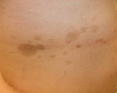
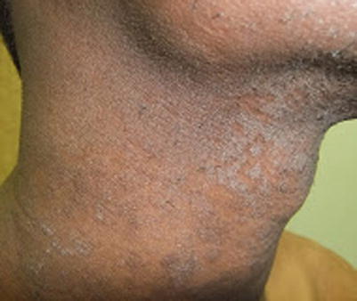
Fig. 16.8
Tinea versicolor. Hyperpigmented patches on the trunk
Fig. 16.9
Tinea versicolor. Extensive case with thin hyperpigmented plaques with fine scale on the neck of a young adult male
Specific Investigations [1, 2]
For diagnosis |
KOH |
Fungal culture |
Histopathology |
For treatment (with systemic antifungals) |
CBC |
LFTs |
KOH preparation shows short fungal hyphae and groups of spores (“spaghetti and meatballs”). Fungal culture is nonspecific for diagnosis and is not recommended, since Malassezia is normal skin flora. Skin biopsy for histopathology with or without PAS is usually not necessary, but is very accurate in confirming the diagnosis, showing abundant organisms in the stratum corneum, associated with no or little inflammation.
Given the very short duration of treatment courses with systemic antifungals, laboratory evaluation is not necessary. In patients with pre-existing hepatic hematologic abnormalities or in patients who require multiple courses treatment for refractory disease, CBC and LFTs should be evaluated.
Management Strategy
In general, topical therapy is the treatment of choice for tinea versicolor. However, a systematic review of controlled trials found most studies to lack the power to detect statistically meaningful differences, although most topical agents are effective compared to placebo. Systemic therapy is highly effective, but is typically reserved for adults with refractory or recurrent disease, and evidence from trials evaluating systemic treatment in children is not yet available. Nonetheless, the most effective regimens based on systematic review of studies in adults are itraconazole (200 mg/day × 5–7 days), fluconazole (300 mg/week × 2 weeks), and pramiconazole (200 mg/day × 2 days) [37, 38].
In studies including both adult and older pediatric patients, ketoconazole cream resulted in 98 % clinical response and 84 % mycologic cure rate compared to placebo. Ketoconazole shampoo was also effective, with clinical response comparable between a single application of ketoconazole shampoo and a 3-day regimen (73 % versus 69 % effective). Topical terbinafine 1 % cream demonstrated mycological clearance in 81 % of patients and clinical response in 72 % of patients with TV. Seventy-seven percent of patients treated with ciclopirox cream, compared with 45 % of patients treated with clotrimazole, had clinical and mycologic cures.
In a randomized, double-blind study, selenium sulfide 2.5 % lotion was superior to placebo; in clinical practice, selenium sulfide 2.5 % shampoo is more often prescribed. Another double-blind study showed zinc pyrithione shampoo was superior to its shampoo base, with all treated patients demonstrating clinical response.
Medication | Dosing | Evidence level |
|---|---|---|
Whitfield ointment (3 % salicylic acid and 6 % benzoic acid) | Twice daily for 4 weeks | A* |
Sulfur-salicylic acid shampoo | Once daily × 1 week | A |
Propylene glycol 50 % in water | Twice daily × 2 weeks | B* |
Selenium sulfide 1 % shampoo (non-prescription) | Once daily × 7 days | E |
While these are not typically used for the treatment of TV, older studies have shown good efficacy. In one double-blind trial, Whitfield ointment was equally effective compared to clotrimazole 1 % cream, producing 80 % mycologic cure rate. Treatment of tinea versicolor with sulfur-salicylic shampoo led to mycologic cure in 86 % of patients. A smaller study of 20 patients using propylene glycol 50 % in water BID reported that all patients demonstrated clinical response, with only 10 % having slight burning sensation after application.
Tinea Nigra
Clinical Features
Tinea nigra is a rare asymptomatic infection of the palms or soles, caused by the dematiaceous fungus Hortaea werneckii. It presents as pigmented macules or patches, generally unilateral in distribution, and is more common in tropical regions [48].
Specific Investigations
For diagnosis |
KOH |
Dermoscopy |
Histopathology |
For treatment |
No specific laboratory investigations required |
Clinical exam and KOH are sufficient for diagnosis in most cases, but the most common clinical concern is acral nevus or melanoma. Dermoscopy has been shown to be a useful non-invasive tool to aid in discrimination from melanocytic lesions and, in one series, was shown to suggest the diagnosis in over 50 % of cases. Dermoscopic findings include superficial fine, light brown strands which form a reticular patch with uniform brown color; unlike melanocytic lesions, furrows and ridges are not followed [49]. Skin biopsy is usually performed to rule out a melanocytic neoplasm, and is definitively diagnostic, demonstrating numerous pigmented hyphae in the stratum corneum.
Management Strategy
Treatment of this superficial mycosis is largely based on case reports, with evidence-based studies and trials with statistical power lacking.
Medication | Dosing | Evidence level |
|---|---|---|
Ciclopirox olamine gel 0.77 % | Three times daily × 3 days | E* |
Medication | Dosing | Evidence level |
|---|---|---|
Isoconazole cream | Twice daily × 20–30 days | E* |
Terbinafine 1 % cream | Daily × 15 days | E* |
Ketoconazole 2 % cream
Related posts:Stay updated, free articles. Join our Telegram channel
Full access? Get Clinical Tree
 Get Clinical Tree app for offline access

|