Fig. 25.1
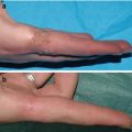
During the first minutes of performing cryosurgery, there is an immediate reaction that is clinically manifest as burning pain of short duration, formation of a white block of ice, and edema. Histologically, this corresponds with vasodilation or dilated blood vessels, congestion (many erythrocytes inside the vessel lumen). Some epithelial cells begin to show changes
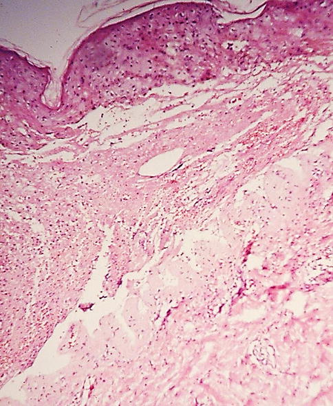
Fig. 25.2
In the first 24 h, a blister formation was clinically observed. Most cases presented serum or blood serum content. Microscopically, epithelial necrosis was seen as well as subepidermal blister formation
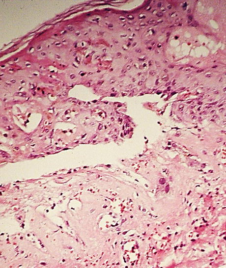
Fig. 25.3
Upon a closer look, necrotic keratinocytes could be seen. An eosinophilic material was present inside the blister as well as inflammatory cells and erythrocytes
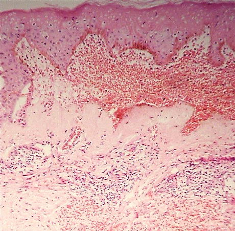
Fig. 25.4
 Preoperative Care for Cryosurgery
 Role of Reflectance Confocal Microscopy in Cryosurgery
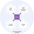 Theoretical Principles of Immunocryosurgery
 In Vivo Reflectance Confocal Microscopy Assessment of Wound Induction and Repair of a Skin Injury Produced by Liquid Nitrogen: An Atlas
 Cryosurgery for Warts
 Cryosurgery for Vascular Lesions


Clinically, some blisters can be hemorrhagic. Histopathologically, one can observe the same subepidermal blister with countless erythrocytes in its cavity and dense lymphocytic inflammatory infiltrate
Related posts:
Preoperative Care for Cryosurgery
Role of Reflectance Confocal Microscopy in Cryosurgery
Theoretical Principles of Immunocryosurgery
In Vivo Reflectance Confocal Microscopy Assessment of Wound Induction and Repair of a Skin Injury Produced by Liquid Nitrogen: An Atlas
Cryosurgery for Warts
Cryosurgery for Vascular Lesions
Stay updated, free articles. Join our Telegram channel
Full access? Get Clinical Tree
