Fig. 17.1
Tibiotalar and subtalar joint dissection after transecting and elevating the fibula preserving distal attachment
Anterior and posterior ligamentous structures are transected from the fibula (Fig. 17.1), preserving soft tissues around the talar neck to preserve vascular supply.
Subtalar joint is accessible after removing fat pad (Fig. 17.1).
17.7 Distal Tibial Reconstruction with Use of a Circular External Fixator and an Intramedullary Nail
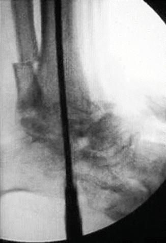
After preparing subtalar and tibiotalar joints, the foot is positioned in correct alignment (neutral to 5° of dorsiflexion in the sagittal plane, 5° of valgus in the frontal plane, and 5° of external rotation in the axial plane) for ankle arthrodesis. A threaded Kirschner wire is inserted through the calcaneus. Entry point of the wire and intramedullary nail is determined with image intensifier as the intersection of joint midline at frontal plane, with anterior 1/3 joint line at sagittal plane. Hole is enlarged with a cannulated drill over Kirschner wire and guidewire is inserted (Fig. 17.2). Reaming is performed over the guide in 0.5 mm increments. Medullary canal has to be reamed 2 mm over the diameter of the nail to be inserted.

Fig. 17.2
Guidewire is inserted and reaming performed
Corticotomy is performed at the planned level with a gigli saw. At least 6–8 cm bone should be preserved proximally.
Intramedullary nail is inserted and nail is locked.
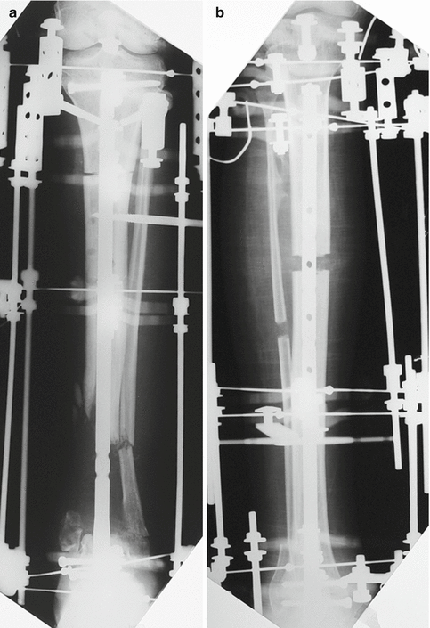
For bone segment transport without lengthening, both ends of the nail are locked (Fig. 17.3a).

Fig. 17.3
(a) In case lengthening over nail is being performed, proximal holes are left unlocked. (b) For segment transport, both holes have to be locked
For pure lengthening, just the distal end of the nail is locked (Fig. 17.3b).
Due to its proximal sagittal curvature (distal for this case as it is delivered retrograde), tibial intramedullary nail is preferred, which provides a dorsiflexion or plantar flexion effect to the ankle.
Circular external fixator is prepared according to the procedure. For lengthening, one ring is placed proximal to the osteotomy and two rings distally (Fig. 17.3a). Proximal two rings are used for lengthening, while the distal ring and the foot ring compress the arthrodesis site.
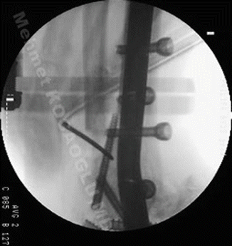
Usually, there is enough bone present at the proximal tibia, just above the tip of the nail for the fixation. Both Schanz screws and Kirschner wires can be used. Rest of the fixation is achieved with wires, passing at least 1 mm away from intramedullary nail (Fig. 17.4). Foot ring is adapted with calcaneal Schanz screws or wires and metatarsal wires.


