Fig. 19.1
Hidradenocarcinoma. A reddish nodule on the scalp. The diagnosis is a histological one
Pathology
The tumor is characterized by a dermal nodular or nodulocystic infiltrative pattern of growth composed of epithelial sheets with a typical clear cell change (Fig. 19.2). The epithelial cells may have also a squamoid or basaloid appearance. Rarely, mucinous differentiation is evident. Central necrosis within lobular epithelial aggregates is sometimes present (comedo variant) (Fig. 19.3). A characteristic feature is the presence of well-developed ducts (Fig. 19.4). Although the tumor may have a deceptively bland appearance, the cells show mitotic activity and nuclear pleomorphism. Perineural infiltration may be found (Fig. 19.5). Pagetoid cells can be identified in the overlying epidermis. The tumor stains with antibodies to keratin AE1/3, cytokeratin 5/6, Ki67, and p53 and may be positive for CEA, EMA, and GCDFP-15. Moreover, like other skin tumors with eccrine differentiation, it also expresses positive staining for estrogen and progesterone receptors. Rarely, hidradenocarcinomas show a t(11;19) translocation and the amplification of the Her2/neu gene.
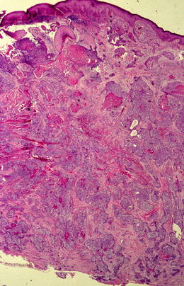
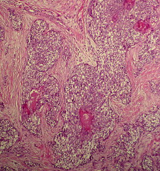
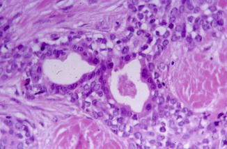
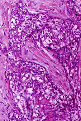
Fig. 19.2
A nodular dermal growth composed of epithelial sheets with squamoid areas and ductal differentiation
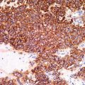
Fig. 19.3
Islands of epithelial sheets with a typical clear cell change, squamoid differentiation and central necrosis
Fig. 19.4
A characteristic feature is the presence of well-developed ducts
Fig. 19.5


Perineural infiltration may be found. Note clear cell change
Related posts:


Stay updated, free articles. Join our Telegram channel
Full access? Get Clinical Tree
