Fig. 35.1
Design of the flap on the donor face

Fig. 35.2
After the cervical midline incision was performed: (a) Submandibular gland was isolated and excised, (b) sternocleidomastoid and omohyoid muscles were removed, (c) carotid sheath was explored
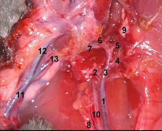
The sternocleidomastoid muscle was detached from its insertions and removed, exposing the omohyoid muscle, which was excised allowing the visualization of the carotid sheath (Fig. 35.2c). The common carotid artery was separated from the vagus nerve and internal jugular vein. For better visualization of the external carotid artery and its branches, the posterior belly of the digastric muscle was removed and the greater horn of the hyoid bone was cut. Internal carotid artery and branches of external carotid artery were fully exposed (Fig. 35.3). The internal carotid artery and all branches of the external carotid artery (superior thyroid artery, ascending pharyngeal artery, lingual artery, ascending palatine artery, facial artery and internal maxillary artery) with exception of the superficial temporal artery were ligated. The dissection of the hemiface flap was carried out above the masseter muscle elevating the branches of facial nerve with the flap. The parotid gland was included in the flap. In the retromandibular region, the maxillary vein and the vein draining the pterygoid plexus were ligated. The external auditory canal and the facial nerve were cut at the osteo-cartilaginous junction and stylomastoid foramen respectively. At this point the cranial incision was performed. The periosteum was incised and the cranial soft tissues were raised subperiosteally up to the temporal line. Thereafter, the dissection was continued in a subfascial plan. The upper and lower eyelids were not included in the flap. At the base of the neck, the flap was raised above the cervical muscles. After transaction of platysma and levator auris longus muscles, the flap was elevated above trapezius up to the external auditory canal.


Fig. 35.3
Omohyoid and digastric muscles, hyoid bone and glossopharyngeal nerve were removed to expose the external carotid artery and its branches. 1 Common carotid artery, 2 internal carotid artery, 3 external carotid artery, 4 superior thyroid artery, 5 lingual artery, 6 facial artery, 7 superficial temporal artery, 8 internal jugular vein, 9 glossopharyngeal nerve, 10 vagal nerve, 11 external jugular vein, 12 linguofacial vein, 13 maxillary vein
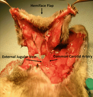
The completely raised hemifacial allograft was pedicled on the common carotid artery and external jugular vein (Fig. 35.4). The hemiface flap was included the ear, parotid gland, facial nerve, external lacrimal gland, external auditory canal, lymph nodes and most of the hemifacial skin. Flap was left pedicled until the preparation of the recipient was completed.


Fig. 35.4
The hemiface flap was elevated above the muscles based on common carotid artery and external jugular vein
Preparation of the Recipient
The skin on the same side of the recipient face was marked as previously described for the donor and facial skin was removed as a full thickness skin graft (Fig. 35.5


Stay updated, free articles. Join our Telegram channel

Full access? Get Clinical Tree


