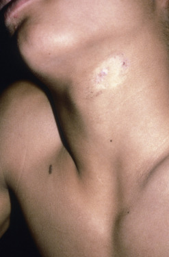
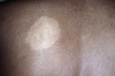
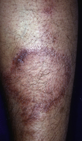
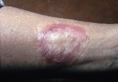
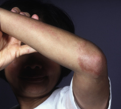
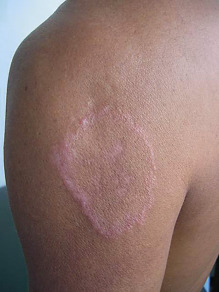
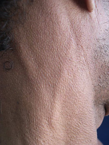
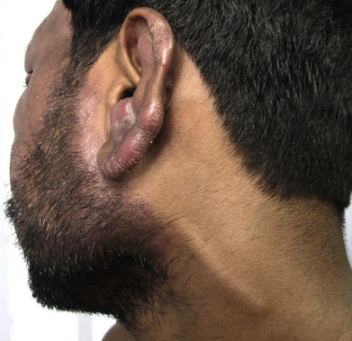
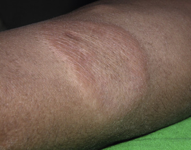
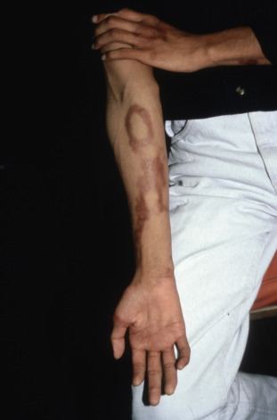
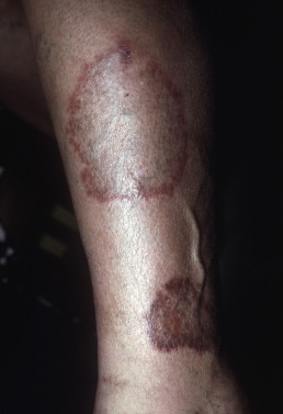
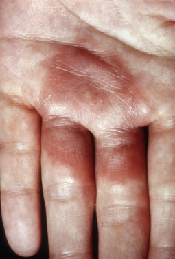
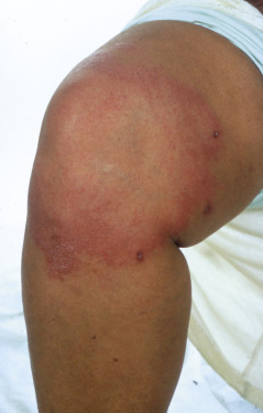
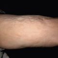
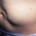
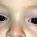
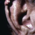
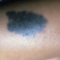
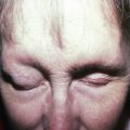
Clinical manifestations of leprosy vary from the subtle hypopigmented patches of indeterminate leprosy to the diffuse facial infiltration and nodules of lepromatous leprosy. The clinical and histologic manifestations of the disease are a reflection of the immune response and bacterial burden. Indeterminate leprosy demonstrates sparse perivascular lymphoid infiltrates and onion skin fibrosis as a manifestation of chronicity. The clinical findings are also subtle with little to no erythema or induration. Tuberculoid leprosy presents as indurated erythematous anesthetic plaques; borderline disease is characterized by annular erythematous indurated lesions, and the lepromatous pole by papules, nodules, and diffuse induration with loss of lateral eyebrows and progression to leonine facies. Diffuse induration correlates with a diffuse dermal histiocytic infiltrate. Globi (clusters of organisms) are easily identified in tissue sections.
Reactional states include reversal reactions characterized by increasing induration, pain, and neurologic manifestations as a reflection of a heightened cell-mediated immune response. In contrast, erythema nodosum leprosum represents a reaction to locally formed immune complexes and is characterized histologically by leukocytoclastic vasculitis in areas of high bacterial burden. Lucio phenomenon demonstrates thrombosis of large and small vessels with variable vasculitis and presents with stellate ulcers and retiform purpura on a background of indurated skin. This portion of the atlas will guide you through the various clinical presentations of Hansen disease.

Related posts:
Stay updated, free articles. Join our Telegram channel

Full access? Get Clinical Tree


