1. Vascularity of the recipient site
2. Single port micro-harvesting cannulas (16G/2 mm or less)
3. Long suctioned tubular passageways
4. Avoidance of splash injuries
5. Low pressure vacuum (18 mm) exposure and ultraviolet ray exposure
6. Limited exposure to non-isotonic solutions,
7. Avoidance of scraping and stirring of the tissue
8. Elimination of centrifugal force
9. Limited exposure to lidociane, epinephrine, and triglycerides
10. Absence of sepsis
11. Avoidance of decanting
12. Use of a closed system
13. Multi-layered droplet injection with sufficient parcel size
14. Sufficient nourishment for transferred fat parcels
In order to maximize graft survival and mitigate against potential assaults to the optimal survival of the adipocyte, harvesting techniques should be adjusted accordingly [2]. Additionally, fat storage utilizing accredited fat banking facilities, such as American CryoStem and BioLife Cell Bank, and AdiCyte is relatively inexpensive and can be used by patients for long-term storage of their harvested fat tissues. This allows for serial transfer to the hands or to other sites permitting further retention and stem cell amplification. Slow freeze methods are more strongly correlated with survival than other storage techniques. Every patient should be offered the opportunity to store fat. There is no correlation between fat survival and overcorrection. However, overcorrection to the point of creating high intra-graft pressure may significantly decrease graft survival as a result of decreasing blood flow and restricting adequate nutrition and oxygenation required for optimal graft survival.
48.2 Aesthetics of the Aging Hand
After the face, the hands are the second most visible telltale sign of one’s age. Looking younger after facial rejuvenation treatments or procedures, such as injectables, neurotoxins, topical treatments and peels, laser treatments, facelifts, or eyelid surgeries, can conflict with untreated aged-appearing hands that aesthetically do not match the face. The hands may give away age more than the face, especially if the face has been medically rejuvenated, and the hands have not. The most significant factors that lead to this hand-aging discrepancy are noted in Table 48.2.
Table 48.2
Hand-aging factors
1. Loss of fullness |
2. Presence of wrinkles and veins |
3. Prominent tendons and joints |
4. Thin skin |
5. Contour deformity |
6. Spots |
7. Absence of nail polish and jewelry |
Fat transfer can be used to effectively fill up hands and reduce the visibility of veins and tendons [3], whereas the function of the hands is clearly primary to their appearance; to many patients, aging-associated changes of the hands are of major importance (Fig. 48.1) [3]. There are many factors that characterize the appearance of aging of the hands, including wrinkle formation as a result of dermal and subcutaneous volume loss, increased visibility of tendons and veins, age spots, and precancerous lesions.
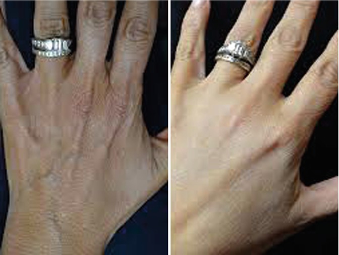
Fig. 48.1
(Left) Hand appearance before fat transfer. (Right) Hand shows reversal of aged appearance following fat transfer
All of these changes are part of a three-dimensional aging process that involves osseous and subcutaneous structures as well as the skin. Therefore, they cannot be fully addressed by the majority of treatment options, such as topical treatments and lasers [4, 5]. The usage of a validated hand grading scale to objectively quantify the severity of hand aging and to maintain the reliability of evaluation, diagnosis for clinical practice, and research is fundamental. Validation grading scales, such as the Five-Point Photonumeric Scale, of Carruthers et al., or the Merz Validated Hand Grading Scale, are examples that can be used in clinical practice to objectify and document the assessment of the aging hand and should be used on a consistent basis for patient evaluation and documentation [6].
48.3 Treatment of Aging Hands
Although there are multiple options available for three-dimensional rejuvenation of aging hands, no one treatment modality alone will necessarily address all of the factors that influence the aesthetic perception of aging hands. A comprehensive approach to hand rejuvenation includes both cutaneous rejuvenation and laser therapies using modalities listed in Table 48.3. Sclerotherapy may be used effectively to treat many cases of excessively prominent veins of the aging hand (Fig. 48.2).
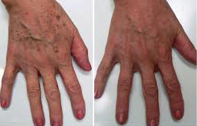
Table 48.3
Cutaneous and laser modalities
1. Topical medical grade skin care |
2. Microdermabrasion |
3. Chemical peeling |
4. Intense pulsed light sources |
5. LED devices |
6. Laser therapies |
(a) Pigment lasers |
(b) Skin tightening lasers, |
(c) Oblateiv non-oblateiv resurfacing |
Fig. 48.2
(Left) Hand before fat transfer. (Right) Hand after fat transfer with residual prominent veins
Certain newer dermal fillers and collagen stimulators, such as hyaluronic acid fillers, poly-l-lactic acid (PLLA), PMMA, and calcium hydroxylapatite, can be selectively used under proper indications to improve the appearance of soft tissue atrophy of the hands [7]. However, soft tissue atrophy of the aging hand is best treated with autologous fat augmentation. Currently, the ability to enrich autologous fat grafts using platelet-rich plasma (PRP) and/or regenerative cells which have been characterized as stem cells, stromal vascular fraction (SVF), mesenchymal cellular extracts (MCE), adipose-derived regenerative cells (ADRC), and bone marrow-derived regenerative cells (BMDRC) provide an exciting opportunity to improve the ultimate retention of these fat grafts and superior outcomes of the fat augmentation procedure.
48.4 Harvesting
Various harvesting techniques have been described and are used to obtain adipose tissue for injection. These include, but are not limited to, the Coleman Method, the Simple Spring Syringe Method, the Manual Syringe Method, the Tulip Syringe, LipiVage, Mega-Trans, Body-Jet, Vaser, and Nutational Infrasonic liposuction. There is an absence of standardized studies to confirm which technique of harvesting and injecting provides for the greatest average percentage of retained volume. A basic list of required equipment and supplies for harvesting is found in Table 48.4. In addition, a list of other basic disposable/products can be found in Table 48.5.
Table 48.4
Equipment and supplies for harvesting
Required | Optional |
|---|---|
Procedure table with arm rest | Mayo stand and back table |
Back table or mayo stand | Clicker lock |
Harvesting cannulas | Syringe stands |
Transfer cannulas | Luerlock hubs |
Basic liposuction cannulas | EKG |
Autoclave w/dry cycle | Crash cart |
Sink (to scrub and clean instruments) | SCD’s |
Cannula cleaning brushes, syringes | Fluid warmer |
Protein denaturing solutions (enzymatic cleaner) | Infiltration and aspiration pump |
Infusion pump: | |
Biohazardous waste | (a) Infusion cannula |
Management | (b) Tubing |
Terminal cleaning materials/supplies | Aspiration Pump |
(a) Tubing | |
(b) Canisters | |
(c) Rack | |
(d) BP monitor | |
(e) Oxygen | |
(f) Pulse oximeter |
Table 48.5
Disposables/products
Non-steriled objects | Sterile procedure items | |
|---|---|---|
Pens for marketing pt | Bed cover | |
LR or saline (1,000 ml bags) | Mayo stand cover or back table cover | |
Medications (lidocaine, epi, NaBicarb, PO, pre/post) | Sterile ½ sheets | |
Sterile gloves and gown | Blade plus handle | |
Hat, mask, shoe covers | Syringe + needles (drawing and delivering) | |
Canisters | Basins | |
Garment (possible patient-supplied) | Betadine gel prep | |
Blue towels | ||
Sterile marketing pen | ||
Terminal cleaning supplies | ||
Biohazard bags (+ mechanism for disposal) | ||
Enzymatic cleaner | ||
Shoe covers | ||
Disposable underwear for pt. | ||
Absorbent pads (e.g., Chux) | ||
Steristrips | ||
Foam | ||
Drains | ||
Towel clamps | ||
Blankets for patient | Sterile tubing (infusion and aspiration) | |
Attention should be focused to ensure that all required pre-procedure preparations, including normal pre-liposuction standard of care requirements, are met. Adequate analgesia should be provided prior to, during, and post procedure. As a stand-alone procedure, harvesting an adequate amount of fat for the purposes of transfer to the hands should, under almost all circumstances, require no more than mild oral sedation and local tumescent anesthesia. The total amount of Klein’s solution wherein the concentration of lidocaine is 100 mg per 1,000 mL of normal saline should not generally need to exceed 250 mL. Whereas the volume of fat required for non-enriched grafting to the hands is in the range of 15–20 mL of fat per hand, the addition of cell enrichment with SVF requires an additional volume of up to 60–100 mL of fat for SVF processing. Therefore, the total volume of adipose tissue required for SVF-enriched fat grafts to the hands will generally be in the range of 90–140 mL.
To date, limited clinical studies are available, with only some reaching a level of evidence to definitively indicate the most superior methods of achieving maximum graft retention. Additionally, the current consensus is that tumor induction by autologous fat grafting is unproven and that the risk associated with autologous fat transplantation is considered to be minor [8
 Stem Cell Applications: An Overview
 The Adipose Organ: Morphological Perspectives of Adipose Tissues
 The Role of Adipose Tissue-Derived Stem Cells and of Angiogenesis
 Advantages of the Transplantation of Fat in Plastic and Reconstructive Surgery
 Tuberous Breast Correction with Stem Cell Fat Transfer
 Stem Cell Fat Transfer for Mastoplasty Using VASER™ Ultrasound and Office-Devised Stem Cell


Related posts:
Stem Cell Applications: An Overview
The Adipose Organ: Morphological Perspectives of Adipose Tissues
The Role of Adipose Tissue-Derived Stem Cells and of Angiogenesis
Advantages of the Transplantation of Fat in Plastic and Reconstructive Surgery
Tuberous Breast Correction with Stem Cell Fat Transfer
Stem Cell Fat Transfer for Mastoplasty Using VASER™ Ultrasound and Office-Devised Stem Cell
Stay updated, free articles. Join our Telegram channel
Full access? Get Clinical Tree
