Hair and Scalp Disorders Resulting in Hair Loss
Herbert P. Goodheart
Hendrik Uyttendaele
 Androgenic alopecia
 Alopecia areata
 Diffuse alopecia
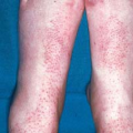
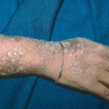
Telogen effluvium
Anagen effluvium
Senescent alopecia
 Scarring alopecia
Chronic cutaneous lupus erythematosus
Lichen planopilaris
Central centrifugal cicatricial alopecia
Traction alopecia
Sarcoidosis
Folliculitis decalvans
 Pseudofolliculitis barbae and acne keloidalis
Overview
Hair has great social and cultural significance in all human societies. It is found on most areas of the human body, except on the palms of the hands, the soles of the feet, and mucous membranes.
Types of hair
Lanugo, the fine hair that covers nearly the entire body of fetuses.
Vellus, the short, fine, “peach fuzz” body hair that grows in most places on the body. Vellus hairs are soft and short. It is seen in areas of male pattern baldness.
Terminal, the fully developed hair, which is generally longer, coarser, thicker, and darker than vellus hair and does not appear until puberty.
Hair texture and shape
Hair texture and shape is genetically determined to be straight, curly or wavy, and it can change over time. It can also be affected by hair styling practices such as chemical straighteners, braiding, or curlers.
Whether hair is curly or straight is determined by the shape of the follicle itself and the direction in which each strand grows out of its follicle. For example, curly hair is shaped like an elongated oval and grows at a sharp angle to the scalp (see Ill. 10.1).
Cycles of Hair Growth
Hair grows in long cycles over many months: a growth (anagen) phase (see Ill. 11.1) is followed by degenerative (catagen) phase, then a resting (telogen) phase, with different hairs alternating phases.
Androgenic Alopecia
Hair loss
Some degree of scalp hair loss or thinning generally accompanies aging in both sexes, and it’s estimated that half of all men are affected by male pattern baldness by the time they are 50 years of age.
Drugs used in cancer chemotherapy frequently cause a temporary loss of hair, because they affect all rapidly dividing cells, not just the malignant ones. Certain diseases and traumas can cause temporary or permanent loss of hair (e.g., systemic lupus erythematosus, thyroid disease).
Basics
Androgenic alopecia (AGA), also known as common baldness (male- or female-pattern baldness), is an extremely common, noninflammatory type of alopecia whose incidence increases greatly with advancing age. AGA is not a disease but a normal consequence of aging. In most, if not all, cultures, hair plays a powerful role in a person’s psychosexual identity and self-image. It is not surprising that in our youth- and image-driven society, hair replacement and retention methods have taken on almost the status of a subspecialty in health care.
AGA is seen more frequently in men than it is in women because women’s hair loss tends to be less apparent, is less extensive, and generally begins at a later age than it does in men.
The condition is genetically determined (autosomal dominant with variable penetrance). The incidence and severity of AGA tend to be highest in white men, followed by white women; it is second highest in Asians and African-Americans and lowest in Native Americans and Eskimos.
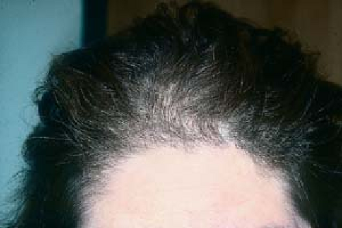
 10.1 Male-pattern alopecia. This is characterized by an M-shaped pattern of hair loss on the front and vertex of the head. |
Pathogenesis
AGA is caused by an androgenic action on hair follicles that shortens the anagen (growth) phase of the hair cycle, thus producing thinner, shorter hairs in a process known as miniaturization.
It occurs through the gradual conversion of terminal hairs into indeterminate hairs and finally to short, wispy, nonpigmented vellus hairs.
In men, this type of alopecia usually begins in late adolescence, with hair loss often starting at the parietal hairline.
In women, the onset is more gradual and the loss of hair is more subtle, and it tends to become obvious later (most often after menopause, but occasionally in the third or fourth decade). This produces a thinning of the hair rather than areas of marked baldness. It is thought that estrogen protects against androgen-mediated miniaturization, which explains both the reduction in severity and the increase in incidence after menopause.
 10.2 Female-pattern alopecia. A midparietal pattern of decreasing hair loss is noted here. The integrity of the frontal hairline is maintained. |
Clinical Manifestations
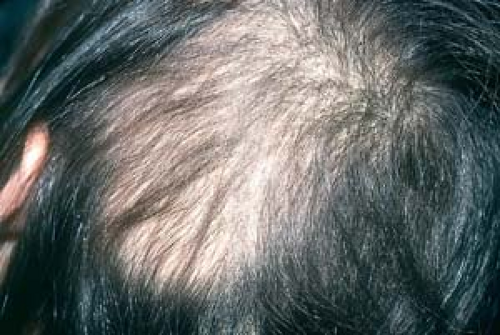
AGA usually produces a patterned type of hair loss. In men and women, hair loss is mostly restricted to the vertex and frontal scalp; hair density on the occipital scalp remains unaffected.
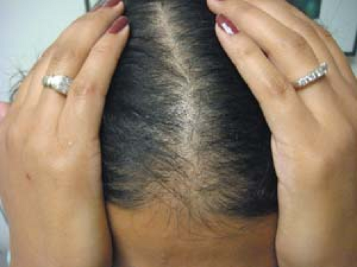 10.3 Female-pattern alopecia. The characteristic “widened part” in a “Christmas-tree” pattern toward the vertex is seen in this patient. |
Description and Distribution of Lesions
Androgenic alopecia produces two typical patterns of hair loss.
In men, the process usually begins in an M-shaped pattern on the front and vertex of the head (this is often referred to as male-pattern baldness).
In women, a thinning of the crown in a “Christmas-tree,” midparietal pattern (female-pattern baldness) is usually noted initially (Fig. 10.3).
Hair loss may progress in both sexes but is often more extensive in men. Thus, in the end stages of androgenic alopecia, many men have only a fringe of remaining hair, whereas women tend to maintain the frontal hairline and do not become frankly bald (see earlier section on “Pathogenesis”).
It should be kept in mind that the following conditions are not only independent causes of hair loss but may coexist and exacerbate AGA; they are discussed later in this chapter.
Telogen effluvium (shedding of resting hairs)
Anagen effluvium (shedding of growing hairs)
Hair loss from thyroid disease
Hair loss caused by iron-deficiency anemia and insufficient calories, protein, or vitamins
Hair loss caused by androgen excess in women
Blood tests and other laboratory studies are necessary only when the diagnosis is in doubt, or other coexisting reasons to explain alopecia are warranted by the history or physical examination (see later section, “Diffuse Alopecia”).
Diagnosis
The diagnosis of AGA is generally based on the clinical pattern of baldness coupled with an absence of clues pointing to a specific disease that may cause hair loss.
Women
Minoxidil (2% solution, applied twice daily) may reduce shedding and may possibly contribute to some regrowth. (The 5% solution of minoxidil may be more effective, but it has not yet been approved for use in women.) The mechanism of action is unknown; however, minoxidil appears to lengthen the duration of the anagen phase, and it may increase the blood supply to the hair follicle. Regrowth is more pronounced at the vertex than in the frontal areas and may not be noted for at least 4 months. Continuing topical treatment with the drug is necessary indefinitely because discontinuation of treatment produces a rapid reversion to the pretreatment balding pattern. The incidence of facial hair growth appears to be increased with the use of the higher-concentration formulation. Patients who respond best to this drug are those who have a recent onset of AGA and small areas of hair loss. In general, women respond better to topical minoxidil than men.
Women with excess androgen may benefit from systemic antiandrogen therapy with agents such as spironolactone, flutamide, or oral contraceptives that decrease ovarian and adrenal androgen production, especially agents that contain a nonandrogenic progestin.
Men
Minoxidil 5% solution and foam (Rogaine), applied twice daily, may reduce shedding and may possibly contribute to some regrowth.
Finasteride (Propecia), 1 mg/day, is an antiandrogen that acts by inhibiting type II 5-alpha reductase, the enzyme that converts testosterone to dihydrotestosterone.
In much higher doses, finasteride is used as a treatment for benign prostatic hyperplasia and prostate cancer.
Recognized side effects include erectile dysfunction and, less often, gynecomastia. It is teratogenic and has not been approved by the U.S. Food and Drug Administration for the treatment of AGA in women.
Men and Women
Hair transplantation is performed by harvesting hairs from donor sites such as the occipital scalp. Hair transplantation, which uses a micrografting technique in which a small incision is used to insert one or more donor hairs, is particularly effective in women, because unlike men, women rarely become completely bald.
A patient’s anxiety regarding hair loss should be taken seriously by his or her health care provider. Hair loss should not simply be “brushed off” as an insignificant cosmetic complaint. The time spent listening to the patient may be helpful in uncovering other emotional or physical problems.
Evaluation of a female patient with AGA may include a complete blood count and testing of thyroid-stimulating hormone and serum iron levels.
Women with symptoms or signs of virilization should undergo a careful history and evaluation for an androgen-excess syndrome. These patients require hormonal studies and may need referral to an endocrinologist.
AGA is very common; therefore, it may coexist with other forms of hair loss. Consequently, a search for treatable causes of telogen effluvium (e.g., anemia, hypothyroidism), especially in patients with an abrupt onset or a rapid progression of their disease, is indicated.
Alopecia Areata
Basics
A common, noninflammatory, idiopathic disorder, alopecia areata (AA) is characterized by well-circumscribed round or oval areas of nonscarring hair loss. Alopecia totalis is a loss of all or almost all scalp hair and eyebrows. Alopecia universalis refers to a total loss of body hair.
AA most commonly affects young adults and children. Occasionally, a family history of AA exists; often, onset is attributed to recent stress or a major life crisis.
 10.4 Alopecia areata. The hair is lost in a round patch. Note the absence of scales or inflammation. |
Pathogenesis
The origin of AA is generally considered autoimmune, because biopsy findings demonstrate T-cell infiltrates surrounding the hair follicles and because AA is sometimes associated with other putative autoimmune disorders, such as the following:
Vitiligo
Thyroid disease (Hashimoto’s disease)
Pernicious anemia
Description of Lesions
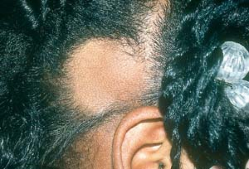
AA most commonly presents as oval, round, or geometric patches of alopecia (Fig. 10.4).
On occasion, a hand lens may reveal tiny “exclamation mark” hairs at the periphery of lesions.
Increased friction (not the expected smoothness) is felt on palpation of lesional skin because of the loss of vellus hairs (Fig. 10.5).
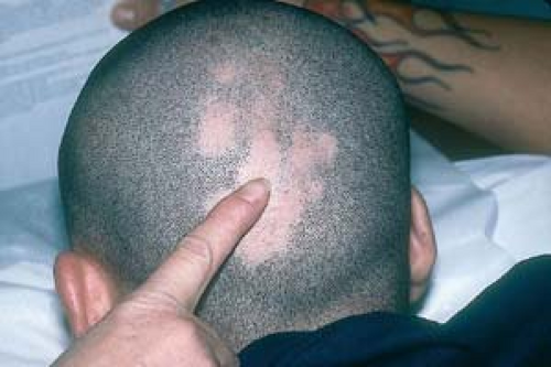 10.5 Alopecia areata. Increased friction is noted on palpation of lesional skin as a result of the loss of vellus hairs. |
Distribution of Lesions
Lesions are most often found on the scalp, eyebrows, eyelashes, and areas of the face that bear hair, such as the beard (Fig. 10.6) or mustache on men.
The entire scalp (alopecia totalis) may rarely be involved, or even the entire body (alopecia universalis), including pubic, axillary, and nasal hair (Fig. 10.7).
Infrequently, nails may demonstrate a characteristic pitting (“railroad tracks”).
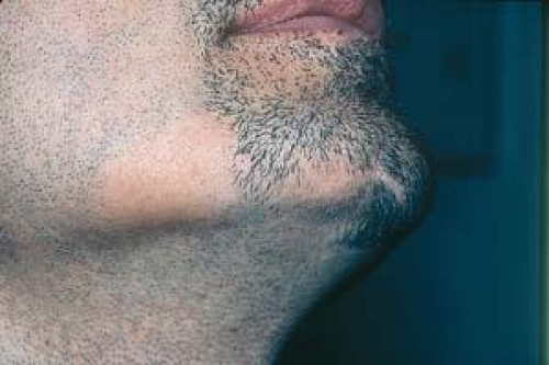 10.6 Alopecia areata. This man’s AA is limited to his beard. |
Clinical Manifestations
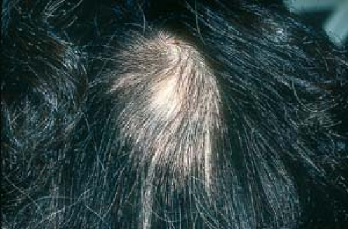
There is usually asymptomatic shedding of hair, which is often discovered by the patient’s hairdresser or a family member.
Frequently, hair spontaneously regrows; however, a recurrence of hair loss may be seen in 30% of patients who had experienced regrowth. Regrowing hair is initially thin and sometimes white (vitiliginous) (Fig. 10.8).
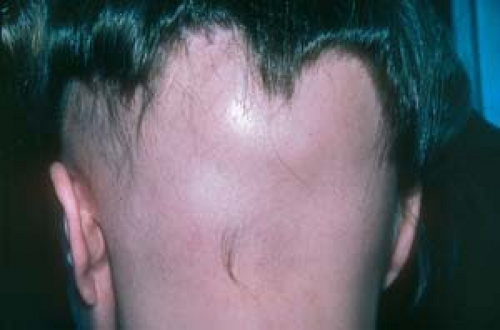
A poorer prognosis is associated with extensive alopecia, an atopic history, and chronicity. Also, when bands of alopecia occur along the hairline margins (ophiasis), (Fig. 10.9) that partially or completely encircle the head, a poorer prognosis is also probable.
Both alopecia universalis and alopecia totalis are generally refractory to therapy and usually last a lifetime; spontaneous regrowth is rare.
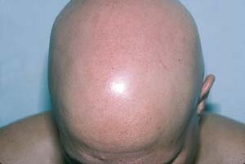 10.7 Alopecia areata, alopecia universalis. This patient has lost all of his hair. He has no eyelashes, intranasal hair, pubic hair, or axillary hair; he also has no hair on his extremities. |
Diagnosis
The diagnosis of AA is generally based on its clinical appearance; however, a scalp biopsy may be performed if the diagnosis is in doubt.
A potassium hydroxide (KOH) test and fungal culture to rule out tinea capitis is negative.
 10.8 Alopecia areata, regrowing hair. In this patient with AA, clusters of hair regrew after intralesional triamcinolone acetonide injections. Some of the regrown hairs are white (vitiliginous). |
 10.9 Alopecia areata, ophiasis pattern. A band of alopecia occurs along the hairline margins encircling the head. |
Tinea Capitis
See Chapter 7, “Superficial Fungal Infections.”
This is seen most frequently in African-American children and uncommonly in African-American adults.
The scalp is often scaly, itchy, and inflamed.
The diagnosis is confirmed when the KOH examination is positive for hyphae or when a fungus grows on Sabouraud medium.
Telogen Effluvium
See also later section.
The hair loss is diffuse.
There is often a history of antecedent illness or childbirth, for example.
Traction Alopecia and Hot-Comb Alopecia
See later discussion.
Other Diagnoses
Trichotillomania
Trichotillomania is also known as compulsive hair pulling (Fig. 10.10).
This is seen most often in young girls.
Hairs tend to be broken at different lengths.
There is an asymmetric loss of scalp hair.
Secondary Syphilis
Secondary syphilis should always be considered in cases of unexplained patchy hair loss (see Fig. 19.19).
The hair loss is referred to as “moth-eaten” in appearance.
Serologic tests for syphilis are generally reactive.
 10.10 Trichotillomania. This condition is seen most often in young girls. Hairs tend to be broken at different lengths. The areas of alopecia are not completely devoid of hair. |
Because mild cases of AA often show spontaneous regrowth, therapy is often unnecessary.
The daily application of superpotent topical steroids, such as clobetasol cream 0.05%, may speed hair regrowth. To increase drug penetration, potent topical steroids, such as fluocinonide cream 0.05%, may be applied, occluded with a plastic shower cap, and left on overnight.
If necessary, intralesional steroid injections into the alopecic patches with triamcinolone acetonide may be administered every 6 to 8 weeks.
Further Treatment Modalities
The numerous treatment modalities for severe extensive AA that have been tried over the years reflect the fact that few are very effective. The success rates associated with the following measures have ranged from no response to varying degrees of partial success:
Irritant therapy involves using a topical anthralin (a coal tar derivative) preparation.Related posts:

Stay updated, free articles. Join our Telegram channel

Full access? Get Clinical Tree