(1)
Professor of Plastic Surgery, Director of Diabetic Wound Center, Director of Cell Therapy Laboratory, Korea University College of Medicine and Korea University Guro Hospital, Seoul, Republic of Korea (South Korea)
Abstract
Many of the chronic wounds are delayed or fail to heal through conventional treatment because attenuated activities of cells responsible for wound healing contribute to the impairment of tissue restoration. The orderly and efficient progression of the wound healing sequence is orchestrated by intercellular communication, much of which is regulated by growth factors and cytokines. Consequently, there is a need for more effective therapy that will stimulate cellular activity of chronic wound patients. Recently, there has been much interest in treating these ulcers with various types of treatment modalities to stimulate cellular functions of chronic wound patients, including growth factors and cell therapies. It has long been hypothesized that using growth factors can promote healing of chronic wounds. Research is currently under way on methods to externally deliver growth factors depleted by diminished cell function in chronic wounds which would therefore facilitate healing; some of these are already being used in clinical settings. Products containing platelet-derived growth factor (PDGF), basic fibroblast growth factor (bFGF), and epidermal growth factor (EGF) for topical use on diabetic foot ulcers are some examples. In this chapter, various growth factor therapies that are used clinically to increase cell activities are described.
Keywords
Growth factorPDGFbFGFEGFOverview
Growth factors are proteins with a molecular weight of 4,000–60,000 Da that can promote cell proliferation and extracellular matrix synthesis; in cellular experiments, they display potent effects in amounts so minuscule that it is measured in pictograms. Growth factors are also commonly referred to by the misnomer “cytokines”; unlike growth factors, cytokines are substances that stimulate cells in any way – not necessarily limited to proliferation, division, and extracellular matrix synthesis.
The names given to growth factors can create confusion. Primarily, growth factors are usually named after the effects they exert on cells or the cells that secrete them, but later studies may reveal the previous findings as incorrect, or add new findings, resulting in discrepancies between the names and actual effects or origins in several cases. For example, unlike its given name, fibroblast growth factor (FGF) is a potent angiogenic factor; likewise, rather than transforming cells, transforming growth factor (TGF) actually has a pivotal role in preventing normal cells from transforming into cancer cells. Names given to growth factors may also depend on the context of its first discovery. For example, a substance would have been called a growth factor had it been discovered by a biochemist, would be called an interleukin if discovered by an immunologist, and a colony-stimulating factor if discovered by a hematologist. Growth factors affect cellular activities even in miniscule concentrations; this is accomplished by delivering special biochemical messages to specific target cells via specific cell membrane receptors.
Growth Factors and Wound Healing
The organic cooperation of various cells involved in each phase of the wound healing process, which culminate in successful wound healing, is the most mysterious phenomenon. The signaling substances credited for making this miracle possible are growth factors.
Research on growth factors during the last two decades has become a source of hope for both patients with nonhealing wounds stagnated somewhere in the normal wound healing process and patients with abnormal scars formed by excessive collagen accumulation. In several studies, even small amounts of growth factors have been found to effectively control cell proliferation, extracellular matrix synthesis, chemotaxis, and more; animal studies have also confirmed that growth factors can promote and enhance wound healing. Recent advancements in tissue engineering and molecular biology have enabled in vitro large-scale biosynthesis of these growth factors, and some are already being sold and used as commercial products. However, it must be noted that more in vivo studies are needed to confirm the capability of these artificially synthesized growth factors in enhancing wound healing in human bodies. The fact that most established studies were carried out on normal wounds indicates that the effects and safety issues of various growth factors on keloids, hypertrophic scars, and chronic wounds must be verified through further studies.
Wounds heal through the separate, overlapping, and complementary actions of complex cellular and molecular processes. Growth factors deliver signals between cells to stimulate cell proliferation or chemotaxis, ultimately inducing extracellular matrix synthesis and angiogenesis. In the early stages of wound healing, vascular injury promotes a massive influx of platelets into the wound area, which secrete various growth factors such as platelet-derived growth factor (PDGF) and TGF-β. These, in turn, attract macrophages, fibroblasts, vascular endothelial cells, and other cells important in wound healing. In the proliferative phase, vascular endothelial growth factor (VEGF), FGF, PDGF, and TGF-β are secreted, inducing angiogenesis and the synthesis of extracellular matrix components such as proteoglycans, fibronectin, and elastin. The role of growth factors does not end with the completion of wound healing: after, growth factors carry out important roles in tissue preservation and in the maintenance of intercellular signal transduction.
Mechanism of Action
Growth factors induce various aforementioned functions by bonding to specific receptors on the cell membranes of target cells. There are several methods for these growth factors to arrive at their target cells. First is the endocrine mode, in which secreted growth factors enter the bloodstream and migrate toward their target cells. Second is the paracrine mode, in which growth factors move via diffusion toward neighboring target cells that are not far away. The last method is the autocrine mode, in which the target cell is affected by growth factors secreted by itself. Growth factors can also induce different effects according to the target cell type. For example, TGF-β promotes the proliferation of fibroblasts and extracellular matrix components secreted by fibroblasts (such as collagen), but inhibits the proliferation of keratinocytes and endothelial cells. Focusing on this characteristic, research on the utilization of specific antibodies to suppress the negative effects of growth factors is being conducted, with the aim of expressing only the positive effects on wound healing.
Background
As the populations of industrialized countries age and become more sedentary, the prevalence of chronic wounds including diabetic ulcers is increasing dramatically. Chronic or nonhealing wound is a major source of disability, morbidity, and mortality in people with chronic diseases. For instance, approximately 20 % of all people with diabetes who develop lower extremity ulcerations ultimately require amputation. However, chronic ulcers respond poorly to conventional treatments, making them very difficult to manage. The pathophysiology of impaired wound healing is complex. Vascular, neurologic, immunologic, and biochemical abnormalities contribute to the impairment of tissue restoration.
The current standard treatment for chronic wounds consists of debridement, control of infection, pressure relief, and lower extremity revascularization as needed. Debridement of a chronic wound should be carried down to healthy, bleeding tissue. The diagnosis and treatment of infections in the chronic wound can be complex. Chronic wound patients often do not respond to infections with elevated body temperature and/or elevated white blood cell count. Osteomyelitis is a frequent complication of chronic wound infections. Osteomyelitis can be detected by a probing test, radiography, scanning with a radioisotope, or magnetic resonance imaging. Infected ulcers can be treated with topical antimicrobials, systemic antibiotic therapy, and appropriate debridement. Avoidance of pressure or weight bearing is essential in the management of pressure ulcers or diabetic foot ulcers. Pressure should be relieved using optimal therapeutic methods. Management of ischemia is also critical, especially in lower extremity ulcers. Vascular insufficiency should be detected by evaluating pulses in the feet, ankle, or toe brachial indices, arteriographic findings, and transcutaneous oxygen pressures. Vascular surgery or percutaneous transluminal angioplasty should be considered for patients with ischemic feet.
However, many of the chronic wounds are delayed or fail to heal through conventional treatment because attenuated activities of cells responsible for wound healing contribute to the impairment of tissue restoration. The orderly and efficient progression of the wound healing sequence is orchestrated by intercellular communication, much of which is regulated by growth factors and cytokines. Severely impaired activities of cells crucial for wound healing are important factors in non- or delayed-healing wounds.
For example, fibroblasts isolated from diabetic foot ulcers show lower proliferative potential and attenuated growth factor production. Hehenberger et al. reported that fibroblasts derived from diabetic wounds showed impaired cell proliferation by 21–35 % compared to those from nondiabetic wounds. Other authors demonstrated that the levels of EGF, PDGF, and TGF-β1 in diabetic wounds were significantly lower than those in nondiabetic wounds. In an immunostaining test for IGF I, immunolabels were absent in fibroblasts harvested from diabetic ulcers. In contrast, intense staining for IGF I was observed in fibroblasts derived from nondiabetic wounds. In addition, there is an excess of metalloproteinases (MMPs) and a decrease in their natural inhibitors (the tissue inhibitors of metalloproteinases, TIMPs) in diabetic wounds. Lobmann et al. reported that the concentrations of MMP-1, MMP-2, MMP-8, and MMP-9 were increased 65-, 6-, 2-, and 14-fold in biopsies of diabetic foot ulcers compared with those measured in biopsies of nondiabetic wounds, respectively. Furthermore, the expression of TIMP was reduced to half in diabetic wounds compared with lesions of nondiabetic patients. All of these pathologic changes can lead to a breakdown of growth factors and extracellular matrix components.
Consequently, there is a need for more effective therapy that will stimulate cellular activity of chronic wound patients. Recently, there has been much interest in treating these ulcers with various types of treatment modalities to stimulate cellular functions of chronic wound patients, including growth factors and cell therapies.
Evaluation of Cell Activity
When deciding a treatment plan for chronic wound patients, predicting functions of the key cells that induce wound healing is important since most of these patients have attenuated cellular activities mainly due to poor systemic conditions for optimal wound healing. However, there has not been a straightforward method to evaluate cell function with ease.
The author hypothesized that functionally active fibroblasts would secrete more collagen at the time of wound formation to restore injured tissue compared to normal conditions. A study was performed to discover if serum collagen level could be used as a predictor for cellular function in diabetic foot patients. Fifty-seven diabetic foot patients who had similar wound characteristics and baseline data were divided into two groups, healed wound group who showed successful healing within 8 weeks and nonhealed wound group who did not within 12 weeks. Serum collagen levels of the two groups were compared.
The results showed that the serum collagen level was 197.7 ± 86.3 ng/ml in the healed group, which was significantly higher than the normal value (76 ~ 163 ng/ml), and 87.9 ± 28.8 ng/ml in the nonhealed group. Based on these results, it may be recommended that the serum collagen level can predict the cellular functions for wound healing in diabetic ulcer patients. However, further research should be carried out to establish this method.
Commercial Growth Factors
Growth factors are growth-promoting substances that increase or enhance cell differentiation, proliferation, and activity. Growth factors are known to play an important role in every phase of the wound healing process. They initiate their effects by attaching to specific receptors on the cell membranes of target cells, thereby activating signal transduction systems. In humans, wounds treated with certain growth factors improved significantly more than wounds treated with a placebo.
Since the effects of growth factors are pleiotropic, and their manifestations are decreased in chronic wounds, it has long been hypothesized that using growth factors can promote healing of chronic wounds. Research is currently under way on methods to externally deliver growth factors depleted by diminished cell function in chronic wounds which would therefore facilitate healing; some of these are already being used in clinical settings. Products containing PDGF, epidermal growth factor (EGF), and basic FGF (bFGF) for topical use on diabetic foot ulcers are some examples.
However, the use of growth factors to facilitate wound healing is extremely costly and limited to chronic wounds that are recalcitrant to traditional interventions. In addition, it is important to emphasize that the growth factor therapy must be used along with other standard principles of chronic wound management, including debridement, infection control, pressure off-loading, and revascularization. Without adhering to these important principles, addition of an active adjunctive modality is unlikely to result in improved healing in diabetic foot ulcer patients.
In this chapter, various commercial growth factor therapies that are used to increase cell activities are described.
PDGF
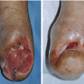
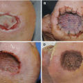
PDGF is a dimer composed of two polypeptide subunits, PDGF A-chain and PDGF B-chain. These subunits form three dimeric forms PDGF-AA, PDGF-AB, and PDGF-BB. PDGF-BB was manufactured by recombinant DNA technology by insertion of the gene for the B-chain of PDGF into the yeast, Saccharomyces cerevisiae (generic name: becaplermin, brand name: Regranex). Becaplermin is a recombinant human PDGF-BB, available as a topical gel. Analysis of healing human wounds showed that becaplermin induces fibroblast proliferation, and differentiation and was found to increase healing in patients with decreased healing capacity, such as people living with diabetes. Becaplermin has a molecular weight of approximately 25 KD and is a homodimer composed of two identical polypeptide chains that are bound together by disulfide bonds. Regranex gel® is a sodium carboxymethylcellulose-based topical gel, containing the active ingredient becaplermin and the inactive ingredients including glacial acetic acid, l-lysine hydrochloride, m-cresol, methylparaben, propylparaben, sodium acetate trihydrate, sodium chloride, and water. PDGF-BB has been sold in the USA, Canada, Europe, and South Korea.
Indication
Becaplermin gel is indicated for the treatment of lower extremity diabetic neuropathic ulcers that extend into the subcutaneous tissue or beyond and have an adequate blood supply. Studies of becaplermin showed that when used with good wound care practices including initial sharp debridement, pressure relief, and infection control, complete healing significantly increased. Pharmacoeconomic studies reinforce the cost-effectiveness of becaplermin as an adjunct to good wound care.
Dosage and Administration
The amount of becaplermin to be applied varies depending upon the size of the ulcer area.
To apply becaplermin gel, proper length of gel is squeezed on to a clean surface, e.g., sterile gauze or wax paper. The becaplermin gel is transferred from the clean surface using an application aid and then spread over the entire wound bed to yield a thin continuous layer. The site of application should then be covered by a saline moistened dressing and left in place for approximately 12 h. The dressing should then be removed and the ulcer rinsed with saline or water to remove residual gel and covered again with a second moist dressing (without becaplermin gel) for the remainder of the day. Becaplermin gel should be applied once daily to the wound until complete healing has occurred. If the wound does not decrease in size by approximately 30 % after 10 weeks of treatment or complete healing has not occurred in 20 weeks, continued treatment with becaplermin gel should be reassessed.
Clinical Trial Study
Many studies compared the efficacy of PDGF-BB with standard wound care in diabetic ulcer patients, and PDGF-BB was found to be more effective than standard therapy in both helping a wound to heal and preventing amputation. However, there was also a published clinical study showing inefficiency of PDGF. Senet et al. compared the healing effect of topical becaplermin gel with hydrogel dressing on hypertensive leg ulcers. Becaplermin gel (0.1 %, in hydrogel) or hydrogel dressing was applied, both in doses of 1 cm/cm2, once daily for 8 weeks. Although complete wound closure rates were comparable after 8 weeks for becaplermin (5 of 28 patients) and hydrogel (3 of 31 patients), no statistically significant differences were observed between the 2 groups at week 12. They concluded that topical becaplermin gel is not superior to hydrogel dressing for hypertensive leg ulcer wound closure.
Precautions
The efficacy of becaplermin gel has not been established for the treatment of pressure ulcers and venous stasis ulcers and has not been evaluated for the treatment of diabetic neuropathic ulcers that do not extend through the dermis into subcutaneous tissue or ischemic diabetic ulcers. The effects of becaplermin on exposed joints, tendons, ligaments, and bone have not been established in humans.
Related posts:
Stay updated, free articles. Join our Telegram channel

Full access? Get Clinical Tree


