Key Words
superior gluteal artery perforator flap, SGAP, inferior gluteal artery perforator flap, IGAP, breast reconstruction
Introduction
The advent of gluteal artery perforator (GAP) flaps has served to increase the surgeon’s armamentarium in autologous breast reconstruction. In 1993, Koshima et al. originally described local-regional GAP flaps for repair of sacral pressure sores. Later in 1995, Allen and Tucker described the utility of the free superior gluteal artery perforator (SGAP) flap in breast reconstruction. Even in thin patients, GAP flaps offer an excellent source of autologous tissue. GAP flaps render soft tissue with a high fat-to-skin ratio, which can be favorable in selected patients. Furthermore, abdominally based flaps may be unavailable due to prior surgery or inadequate volume. Hence, GAP flaps have been championed as an alternative to abdominally based free flaps.
In 2004, Allen et al. further refined the inferior gluteal artery perforator (IGAP) flap to be included into the infragluteal crease, resulting in a more cosmetically acceptable surgical footprint. As with any other perforator flap, muscle is spared which decreases the donor-site morbidity leading to a faster recovery. Additionally, the muscle functionality is preserved. Typical pedicle lengths are 7–10 cm in an IGAP and 5–8 cm in an SGAP. Despite the fact that thigh flaps have essentially replaced buttock flaps as the second choice for perforator flap breast reconstruction, reliability of the tissue quality, abundance of volume, and a reasonable pedicle length of GAP flaps make these operations an essential part of a microsurgeon’s armamentarium.
In this chapter, the authors describe the indications and contraindications of GAP flaps for breast reconstruction with emphasis on patient selection, operative techniques, postoperative care, and management of complications.
Indications and Contraindications
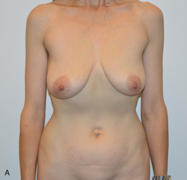
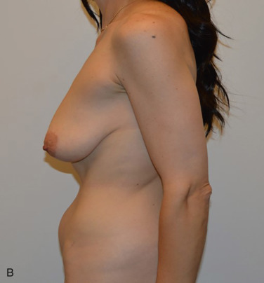
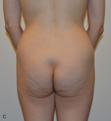
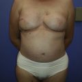
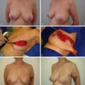
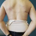
Gluteal artery perforator flaps can be considered in women who have undergone mastectomies and desire autologous tissue reconstruction. Other indications include hypomastia, Poland syndrome, anterior thoracic hypoplasia, and correction of breast asymmetry. Ultimately, it is the patient’s decision as to where she wants the donor-site scar burden to be located. Women who have had surgery that precludes abdominally based reconstruction or have insufficient abdominal tissue are candidates for GAP flap reconstruction. Prior abdominoplasty and extensive liposuction of the abdomen may render the abdomen unusable for perforator flap–based reconstruction. GAP flaps are a logical alternative. Sometimes a patient does not want a long scar that she can see on her lower abdomen, and would prefer a hidden scar in the back ( Fig. 5.1 ).
Liposuction of the buttocks or is a relative contraindication to GAP flap reconstruction. If the area of liposuction was confined to only a portion of the buttock, sometimes a gluteal flap can be designed on a vessel in the non-operated area.
Preoperative Evaluation
As with any surgical procedure, preoperative patient optimization is paramount. It is equally important to manage patient expectations prior to surgery and discuss goals of the surgery, scar burden, possibility of flap complications, and the need for further revisional surgery. A full review in detail of the past surgical history and patient medication list is indispensable as these can potentially preclude surgery.
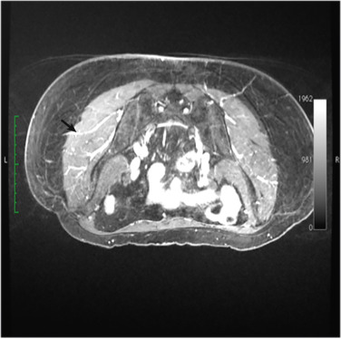
Magnetic resonance angiograms (MRA) are a mainstay of our patients’ preoperative work-up ( Fig. 5.2 ). The angiograms allow a qualitative evaluation of perforator location, course, and caliber. Hence, they are integral to the presurgical planning. Our radiologists use a coordinate system with reference points such as the midine and the gluteal fold to describe the perforators’ location as they exit the muscle. Each perforator coordinate is measured in its vertical and horizontal position with respect to these landmarks. Perforator selection is multifactorial, which is why each case must be tailored according to the imaging and patient body habitus. A large vessel diameter, pedicle of sufficient length for insetting, central location of the vessel on the flap, or a pattern of arborization that suggests perfusion of the tissue to be transferred are all considered favorable for the choice of the pedicle to be used. Use of the angiogram in conjunction with a pencil Doppler facilitates the most accurate identification of the perforators prior to the surgery. This method is particularly helpful in designing the skin paddle that will capture the perforators.
Surgical Techniques
Relevant Anatomy of the Gluteal Region
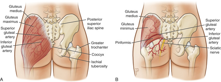
The gluteus maximus muscle measures approximately 24 × 24 cm. It originates from the posterior superior iliac spine, the coccyx, and the lateral aspect of the sacrum. The muscle inserts into the greater trochanter and the iliotibial tract of the fascia lata.
The superior gluteal artery arises from the internal iliac artery and exits through the greater sciatic foramen, above the piriformis muscle perforating the gluteus maximus muscle to supply the overlying skin and fat. The exit point corresponds anatomically to 6 cm below the posterior superior iliac spine and 4.5 cm lateral to the midline of the sacrum. The inferior gluteal artery also arises from the internal iliac artery and along with the sciatic nerve and emerges below the piriformis muscle ( Fig. 5.3 ).
Preoperative Marking
Typical skin paddles range from 6–10 cm in width by 18–22 cm in length. It is incumbent on the surgeon to resect a skin island that will facilitate a tension-free closure. A patient’s preference towards a superior or inferior flap is influenced by the advantages and disadvantages of each gluteal flap procedure. Anatomic considerations are based on a patient’s distribution of fat on the buttock and location of the most favorable vessels in the buttock.
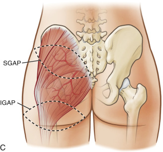
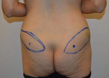
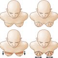
In an SGAP flap, the skin island is elliptical with an oblique axis slanting cephalad from medial to lateral. To limit conformational change, the MRA and the marking should be done in the prone position. The scar of the SGAP can be covered with a bathing suit, however it is located in a more prominent position on the buttock, therefore, harvesting a superior gluteal flap can disturb the superior fullness of the buttock, causing unfavorable flattening of this area ( Fig. 5.4 ).
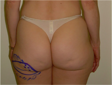
In an IGAP flap, the skin island is elliptical, and the inferior border of the flap should be parallel and placed about 1 cm inferior to the gluteal fold. The fold should be identified with the patient in a standing position. The flap design could be shifted laterally to address “saddlebag” deformities if needed. The scar from an inferior gluteal flap is located in a less prominent area of the buttock in the inferior gluteal crease and can be hidden. The scar is typically visible lateral to the buttock crease, and the natural curve of the inferior buttock crease can be unfavorably flattened ( Fig. 5.5 ).
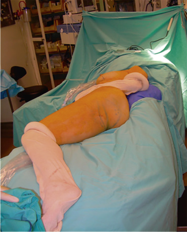
Preoperative Setting and Patient Positioning
For deep venous thrombosis prophylaxis, a subcutaneous heparin injection is given preoperatively and sequential compressive devices are placed on the legs. A prophylactic dose of antibiotic is given, a urinary catheter inserted and general anesthesia given. For a unilateral breast reconstruction, the patient is placed in the lateral decubitus position with appropriate padding of pressure points supported with a bean bag. The upper and lower extremities on the side of the breast reconstruction are prepped in the field. For a unilateral breast reconstruction, a gluteal flap is harvested from the same side of the body as the reconstructed breast. The mastectomy and GAP flap dissection can be performed simultaneously ( Fig. 5.6 ). After the flap is harvested, the patient is placed in supine position for microsurgical anastomosis to the internal mammary vessels and flap insetting. For bilateral simultaneous breast reconstruction, the patient is first placed in a supine position for the mastectomy and preparation of the recipient vessels in the chest. The patient is then placed in the prone position for harvesting of the GAP flaps and closure of the donor sites. The patient is turned again to the supine position for microsurgical anastomosis and flap insetting.
Operative Procedure
A team of two microsurgeons operate simultaneously. The preparation of recipient vessels in the chest and the dissection of the gluteal vessels are performed with loupe magnification.
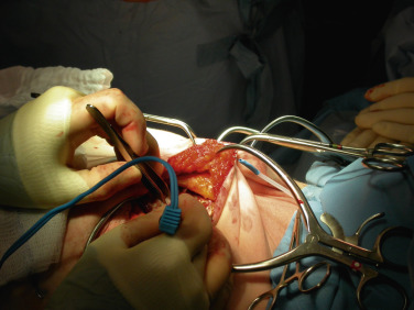
Recipient Site Exposure
The most commonly used recipient vessels are the internal mammary vessels, which run parallel to the sternum, under the ribs ( Fig. 5.7 ). The muscle fibers of the pectoralis muscle are separated at the intercostal space between the second and third or third and fourth ribs. The perichondrium is elevated off the most medial portion of the ribs and the most medial section of the intercostal muscle is resected. A pale yellow layer of fat containing the internal mammary vessels is located under the intercostal muscle and over the pleura. The internal mammary artery is accompanied by one or two veins. The vessels are carefully dissected from the surrounding fat using a bipolar cautery. Side branches of the vein are preserved distally to create a wider lumen in case of size mismatch with the gluteal vein. An intercostal space of 2–3 cm is sufficient to perform the anastomosis. If more space is required, a portion of medial rib cartilage can be removed. The arterial size match is usually favorable if the GAP pedicle dissection was pursued all the way to the source vessel (SGA or IGA). If the pedicle was harvested prior to the larger artery, the pedicle vessel may be of smaller diameter. In this case, a better arterial size match may be achieved by using a perforating artery or a branch off of the internal mammary artery.
Related posts:
 Immediate Implant Breast Reconstruction With Total Muscle Coverage – Two-Stage
 Free Transverse Upper Gracilis Flap Breast Reconstruction
 Partial Breast Reconstruction With Local Tissue Rearrangements
 Immediate Prepectoral Implant Breast Reconstruction
 Fat Grafting for Total Breast Reconstruction
 Partial Breast Reconstruction With Flaps
Stay updated, free articles. Join our Telegram channel

Full access? Get Clinical Tree


