Key Words
conservative breast surgery, reconstruction, local tissue rearrangements, glandular flaps, mastopexy, mammaplasty, local flaps, outcome, surgical planning
Introduction
Breast conservation surgery (BCS) has become an alternative technique to mastectomies as a consequence of its lower morbidity, sensitivity preservation and equivalent overall survival rate. BCS reconstruction consists of a concept that combines BCS with techniques of breast reshaping in order to improve the final aesthetic outcome and to expand the indications for breast conservation without decreasing oncological safety.
Recently, increasing attention has been focused on BCS reconstructive procedures. Among the main techniques, glandular flaps, skin/fat local flaps and mastopexy/mammaplasty are most commonly used with more satisfactory results. The selection of a best technique is determined by the surgeon’s experience and the volume of the BCS defect in relation to the volume of the remaining breast tissue. The main positive aspects of the reconstructive technique should include reproducibility, low interference with the oncologic treatment and long-term results ( Box 16.1 ).
Simple procedure
Less morbidity
Similar skin (color and texture)
Short operative time
Fast recovery
Low interference with the oncologic treatment
This chapter describes local tissue rearrangement (LTR) techniques, which allow the use of remaining breast/local tissue following BCS by glandular flaps, local flaps or glandular reshaping techniques for better aesthetic results. Thorough understanding of these techniques and an adequate consideration of the patient’s breast volume, tumor location, excised volume, and volume of the remaining breast tissue in choosing appropriate surgical techniques will result in a satisfactory outcome. In some cases and depending on the excised volume and the initial breast volume, surgery of the contralateral breast may be necessary to improve symmetry. In addition, the timing of such surgery and the advantages of synchronous versus delayed approaches should also be pointed out.
Indications/Contraindications of LTR
LTR techniques are defined when the BCS defect is reconstructed using one of a range of breast or local flaps, advanced into the lumpectomy defect. Different techniques have been described to repair small or moderate defects. These procedures are all based on advancement or rotation of an area of breast or local tissue to reconstruct the BCS. According to the breast volume, ptosis, and tumor size/location, the patients are evaluated by the plastic surgeon who decides each type of indicated reconstruction. During intraoperative evaluation, the partial breast defects in relation to the initial breast volume, the size and location of the defect, and the amount of tissue available are evaluated and an appropriate procedure can be selected ( Box 16.2 ).
Conservative breast surgery
Unifocal tumors
Small-volume breasts with small resection (type IA)
Medium-volume breasts with small/moderate resection (type IIA and IIB)
Large-volume breasts with small/moderate resection (type IIIA and IIIB)
Free surgical margins (frozen sections)
The advantage of the immediate reconstruction is that the whole procedure can be achieved in a one-stage surgery. Because there is no scar tissue, a tissue rearrangement is easier and the aesthetic outcome can be more predictable. In terms of oncological safety, immediate approach can obtain a widely negative margin around the tumor as a result of wider local tissue excision. This aspect is advantageous and has the potential of reducing the incidence of margin involvement and development of local recurrence. In spite of the advantages, the immediate reconstruction has some negative aspects and can be time-consuming and require specialist training to properly apply these techniques. In addition, radiation therapy usually involves some degree of fibrosis and shrinkage of the remaining breast tissue. Thus, the aesthetic outcome may be unpredictable. Another negative aspect of immediate reconstruction relates to uncertainties about surgical margin involvement and the necessity to return to surgery for re-excision.
Preoperative/Intraoperative Evaluation and Special Considerations
The preoperative evaluation is based on our previously described classification system for breast conservation surgery defects. This system is based on the breast volume, the extent/location of glandular tissue resection, and the remaining available breast tissue. Basically, in patients with small breasts without ptosis and defects that do not cause volume distortion in the breast shape, LTR based on breast flaps can be utilized with a satisfactory outcome. In patients with medium/large-sized breasts, mammaplasty technique is indicated ( Table 16.1 ).
| Type I | Type II | Type III | ||||||
|---|---|---|---|---|---|---|---|---|
| Small volume without ptosis Cup size: A/B | Medium volume with/without ptosis Cup size: C | Large volume with ptosis Cup size: D | ||||||
| A | B | C | A | B | C | A | B | C |
| Small defects Without distortion | Moderate defect Moderate distortion | Large defects Severe distortion | Small defects Without distortion | Moderate defect Small/mod distortion | Large defect Mod/severe distortion | Small defect Without distortion | Moderate defect Small distortion | Severe defect Mod/severe distortion |
Usually, the reconstruction techniques are performed with one of five surgical options: breast tissue advancement flaps, local flaps, bilateral mammaplasty, latissimus dorsi myocutaneous flaps, and complete skin-sparing mastectomy with total reconstruction. The main indications for LTR are for types IA, IIA, and IIIA. In this scenario breast defects are reconstructed with breast tissue advancement flaps in which the defect created is usually spherical or rectangular. The breast tissue is advanced along the chest wall or beneath the breast skin flap to fill the tumor defect.
In type IB, the lateral thoracodorsal flap can be indicated for lateral defects. This flap can be planned as a wedge-shaped triangle located on the lateral aspect of the thorax and then rotated to the breast defect. For small defects, the flap is planned as a triangle located exclusively on the lateral aspect of the thorax. In type IIB, breast defects are most frequently reconstructed with breast tissue advancement flaps and associated mastopexy techniques. The same concept can be applied for types IIIA and IIIB. Defects classified as C (I, II and III) are not adequate to be reconstructed by LTR.
Frequently, type IC defects are converted to a skin-sparing mastectomy and reconstructed with an appropriate technique for each case. Type IIC and IIIC defects should be analyzed individually according to the size of the breast defect in relation to the remaining tissue available. For this purpose, the patient is positioned upright to assess the amount of glandular tissue. Thus, type IIC/IIIC can be subclassified into favorable and unfavorable defects. If there is enough tissue to perform an adequate breast mound shaping, the defect is classified as favorable. For lateral defects, the extended lateral thoracodorsal flap is most commonly used where the inferior and superior limits are designed more obliquely with curved borders to incorporate a large amount of tissue.
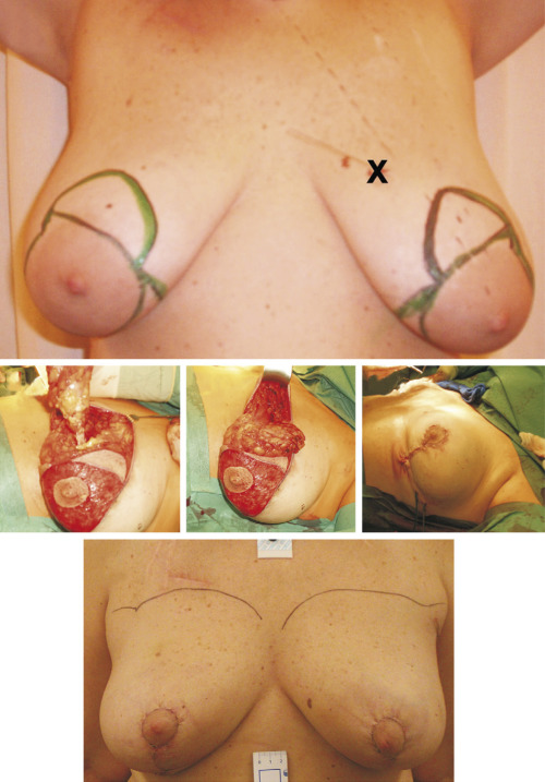
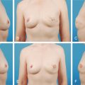
In patients with central/medial defects, the extended latissimus dorsi myocutaneous flap can be used. Conversely, if not enough breast tissue remains, the breast defect is classified as unfavorable and a skin-sparing mastectomy is indicated. In type IIIC defects, sometimes the defect is favorable and the deficiency is most frequently reconstructed with bilateral reduction mammaplasty. A marked reshaping of the breast with available tissue and a similar contralateral breast reduction is then performed ( Fig. 16.1 ).
Surgical Techniques
Relevant Surgical Anatomy
LTRs are based on breast tissue advancement flaps that are supplied by glandular arteries from three main arteries. The great part of the breast tissue extends inferiorly from the level of the second or third rib to the inframammary fold (at the level of the sixth or seventh rib). The breast tissue also extends into the axilla as the glandular tail of Spence, which can be used as a glandular flap. The posterior surface of the breast tissue rests on portions of the fasciae of the pectoralis major, serratus anterior, external abdominal oblique, and rectus abdominis muscles.
The LTA usually arises from the axillary artery and supplies up to 30% of breast blood flow to the lateral and upper outer aspects of the breast. The branches course inferomedially within the subcutaneous tissue to effect anastomoses with branches of the internal mammary artery (IMA) and internal carotid artery (ICA) in the areolar area. The III, IV, and V posterior ICA are the least important arteries supplying the breast.
Preoperative Markings
Despite the simplicity of the LTR techniques, important technical steps must be planned beforehand. Operative planning should include tumor location and the extent of glandular tissue resected, addressing individual reconstructive requirements, thus enabling each patient to receive an individual “custom-made” reconstruction. The success of the procedure depends on patient selection, coordinated planning with the oncological surgeon and careful intraoperative management. In addition, an in-depth dialog concerning alternatives for BCS reconstruction should be undertaken within a multidisciplinary scenario, including the risks and positive aspects of the LTR reconstruction. Usually for any type II or III candidate for immediate BCS, we perform a mammaplasty planning based on classical markings. After the BCS and with an appropriate intraoperative evaluation, we can follow these marks and perform the mammaplasty or, in cases of subtype A, convert to an LTR with breast tissue advancement flaps.
Basic Markings
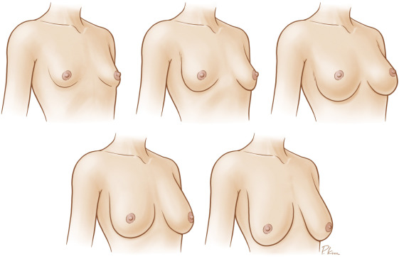
With the patient in an upright standing position, the breasts are examined and the vertical axis as well as the pre-existing inframammary fold are marked. If the breast presents some degree of ptosis, the new nipple–areola complex (NAC) position is defined. For this purpose, the projection of the inframammary fold onto the anterior surface of the breast is drawn and it will indicate the new position of the upper border of the NAC (point A). The new areola is then marked, and a semicircle is drawn. The diameter of this semicircle will vary according to the size of the breast and the amount of breast tissue to be resected. Although the markings for the reduced areola may be made at this point, we usually prefer to perform this intraoperatively. Thus, these markings are not rigid and have to be checked again after the BCS. Points B and C are marked by pinching the skin at the level of the nipple, and seeing the amount of excisable skin in the horizontal plane. Usually, points AB and AC are joined as a curved line, and the transposition of points A, B, and C to the contralateral breast is done with the aid of suture lines placed over the sternal notch and the xiphoid appendix ( Figs. 16.2–16.4 ).
Related posts:
 Immediate Implant Breast Reconstruction With Total Muscle Coverage – Two-Stage
 Free Transverse Upper Gracilis Flap Breast Reconstruction
 Fat Grafting as an Adjunct Procedure in Breast Reconstruction
 Immediate Prepectoral Implant Breast Reconstruction
 Fat Grafting for Total Breast Reconstruction
 Partial Breast Reconstruction With Flaps
Stay updated, free articles. Join our Telegram channel

Full access? Get Clinical Tree


