Fractional resurfacing

CHAPTER 77 Fractional resurfacing


History


Laser skin resurfacing represents one of the least invasive methods with the highest efficacy and safety profile for rejuvenating the aging face. Traditional ablative skin resurfacing rapidly gained early acceptance since the first introduction of the CO2 laser in 1964. Ablative skin resurfacing methods (CO2 or the Erbium:YAG lasers), specifically target intracellular water in the skin based on the theory of selective photothermolysis. The CO2 laser heats the target within cells instantaneously to greater than 100°C leading to vaporization of tissue on the surface layer of the skin; coagulation necrosis of cells and denaturation of extracellular proteins in the next layer; and finally non-fatal cellular damage in the deeper zones of the skin. Ablative lasers remove 100% of the epidermis and varying thickness of underlying dermis that results clinically in a smoother appearance to the skin and skin tightening due to heat induced collagen shrinkage. While ablative lasers have long been the gold standard for the treatment of photodamage, the applicability of the treatment has been limited by the potential for unfavorable side effects and by the prolonged healing period and downtime patients require before returning to their routine. Frequently, patients have post-treatment erythema, edema, burning and crusting. The erythema may last on average 4.5 months and pigmentary alteration, acne flares, herpes infection/reactivation, scars, milia, and dermatitis also occur. Single pass CO2 laser resurfacing can reduce the severity of these side effects, however.


Non-ablative fractional photothermolysis was first introduced in 2005 with the introduction of the 1550-nm erbium-doped fiber laser (Reliant Technologies, Mountain View, CA). Initially the laser was approved for the soft tissue coagulation application, based largely on early studies of forearm tissue. In 2005 Khan et al. reported on the use of the first fractional resurfacing device, a 1550-nm erbium-doped fiber prototype laser system that produced microscopic columns of thermal injury surrounded by intact tissue using a handpiece that scanned across the skin up to 8 cm/second while delivering the microarray pattern to the skin. These microscopic treatment zones (MTZs) range in diameter from 70 to 100 micrometer in diameter and 250 to 800 micrometer in depth and are produced by varying the depth of the focused beam. Tissue in these coagulated zones is not vaporized and the epidermis and stratum corneum are left intact leaving the skin erythematous, edematous, but without evidence of wounding. The device is FDA cleared for the treatment of periorbital rhytids, pigmentary alteration, melasma, skin resurfacing, and surgical scars.


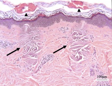
The handpiece of the fractionated laser makes direct contact with the skin’s surface and uses an intelligent optical tracking system to deliver an even array of MTZs. These are columns of tissue coagulation that show no loss in the integrity of the overlying stratum corneum (Fig. 77.1). The incipient wound healing cascade and inflammatory response in which heat shock protein-70 is a central player, initiates a number of incompletely understood events that leads to increase in collagen synthesis and collagen reorganization. Healing also involves the extrusion of damaged epidermal components, termed microepidermal necrotic debris (MEND), which clinically could be observed as a superficial exfoliation following treatment that imparts a fine rough sand paper-like feel to the skin and is associated with a mild bronzing color of the skin in the areas of treatment. This clinically translates into the observed improvement in photodamage, including softening of rhytids, tightening of pore ostia, improvement in dyschromia, and a general textural smoothening of the skin.


Though non-ablative fractional photothermolysis is a well-tolerated and effective modality for an expanding variety of conditions such as photoaging, periorbital wrinkling, mild to moderate acne scarring, melasma, pigmented lesions, and poikiloderma of Civatte, patients generally require multiple treatments to achieve significant results; however even with multiple treatments, severe acne scarring is only minimally improved with non-ablative energies. Additionally, the results for severe photodamage is modest in comparison to fully ablative CO2 resurfacing. Ablative fractional photothermolysis was developed more recently to achieve an improved treatment outcome requiring fewer treatments than non-ablative fractional photothermolysis with shorter downtime and improved side-effect profile than ablative resurfacing.


In early 2008, ablative fractional photothermolysis was introduced in the form of a novel ablative 30W CO2 laser (Reliant Technologies, Inc., Mountain View, CA). Ablative fractional photothermolysis is a technique similar to non-ablative fractional photothermolysis in that a pixilated pattern of microscopic ablative wounds surrounded by healthy tissue is deposited on the skin in a manner similar to that of non-ablative fractional photothermolysis. Ablative fractional photothermolysis combines the increased efficacy of fully ablative techniques with the safety and reduced downtime associated with non-ablative fractional photothermolysis. As the literature on ablative fractional resurfacing is in its infancy and is rapidly evolving at the time of preparation of this chapter, the discussion presented here focuses on non-ablative fractional photothermolysis, and we refer the reader to recent (Chapas et al. 2008) and future publications on this topic.


Physical evaluation


Assessment of patients’ expectations of the procedure needs to be done during the cosmetic consultation. Realistic expectations need to be clearly stated prior to embarking on a series of non-ablative fractional resurfacing treatments. Patients should be advised that only modest results are achieved early in the series, and that more significant improvement will be observed as the series of treatments progress. For the application to photorejuvenation, the optimal candidate would have mild to moderate photodamage. While severe photodamage may be improved as well with fractional resurfacing, the clinical improvement at the endpoint of treatment is expected to be only modest.


The cosmetic consultation visit should focus on the following:


Complete past medical and surgical procedures including complication in the postprocedural periods.


History of hypertrophic scarring and/or keloid formation. While there is a low risk of scarring from fractional resurfacing, a positive history of hypertrophic scarring and/or keloid formation is a marker of a perturbed wounding response and signal that caution should be exercised in treating these patients. Closer follow-up and possibly less aggressive initial treatments should be undertaken until an assessment of an individual patient’s response to therapy can be made.


History of herpes labialis and/or HSV infections elsewhere, use of antivirals and antibiotics in the past, and allergies to these are significant. Patients with active bacterial and viral infections should delay fractional resurfacing treatments until these issues have resolved. Routine use of prophylactic antivirals is recommended for three days starting on the day of treatment.


History of isotretinoin use over last 6 months. These patients should be excluded from treatment because of a theoretical increased risk of scarring.


History of smoking. Patients who smoke generally heal less favorably.


Plans for UV exposure should be gauged before the procedure as UV avoidance is necessary immediately following treatments and during the healing period.


Thorough physical exam noting scars, dyschromia, rhytids, and skin phototype are important considerations in selection of treatment settings for particular applications.


Photography is useful for patient feedback and part of the medical record for later reference.


Mar 4, 2016 | Posted by in Aesthetic plastic surgery | Comments Off on Fractional resurfacing

Full access? Get Clinical Tree

Get Clinical Tree app for offline access