Mastopexy with and without augmentation

CHAPTER 48 Mastopexy with and without augmentation


History


The goal of a mastopexy procedure is to improve breast ptosis by lifting the breast including the gland and the nipple. Aging, gravity, and breast feeding contribute to breast ptosis by stretching the skin and increasing skin redundancy. This may be accompanied by atrophy of the breast gland further exaggerating the ptosis. Correction can be done through a variety of incisions depending on the extent of nipple and/or glandular ptosis. Regnault categorized ptosis by assessing the nipple position relative to the inframammary fold. However, ptosis can be further and more accurately defined by assessing the extent of breast gland below the inframammary fold while also assessing the nipple position on the gland.


Most mastopexy techniques are derived from breast reduction procedures. Breast reduction has a long history dating back to the 1800s. Kraske was the first to reduce the breast and skin horizontally and vertically resulting in the inverted T closure that we continue to use today. Wise developed a template pattern to mark the breast skin that facilitated reliable results with an inverted T closure. Goulian described a mastopexy technique that used a similar template device and inverted T closure, but skin undermining was avoided with the hope of providing a longer lasting result. Whidden developed the tailor-tack mastopexy which used sutures to temporarily reshape the skin envelope prior to marking the breast in order to individualize the skin resection. These early techniques relied on skin to support the breast gland and they were prone to recurrent ptosis. More recent techniques have included glandular reshaping in addition to excision of the redundant skin envelope to produce longer lasting results.


Attempts to minimize scaring led to the development of circumareolar and vertical techniques for mastopexy. The circumareolar technique was first described by Bartels and then popularized by Benelli and Goes. However, this technique often led to a flattened breast with pleating of the skin and stretching of the areola. Spear developed criteria for patient selection for this technique to help limit these complications. The vertical technique was first described by Lassus and then popularized by Lejour. This technique created pillars of breast gland used to reshape and support the breast. Modifications have included a small horizontal skin excision along the inframammary fold as described by Marchac and creation of an inferior glandular flap based on the chest wall used for autoaugmentation as described by Graf and Biggs.


Mastopexy with augmentation in a single stage was first described by Gonzales-Ulloa. Performing a simultaneous augmentation and mastopexy can put the nipple areola complex at risk for vascular compromise. This significant risk has prompted some surgeons to perform this procedure in two stages, especially in cases of severe ptosis such as massive weight loss patients. However, in more moderate cases, mastopexy with augmentation can be performed safely in a single stage. Patients must be aware that there is a high incidence of revision when combining mastopexy with augmentation. Fortunately, adding an implant typically helps fill the skin envelope, reducing the extent of mastopexy required.


Preoperative assessment
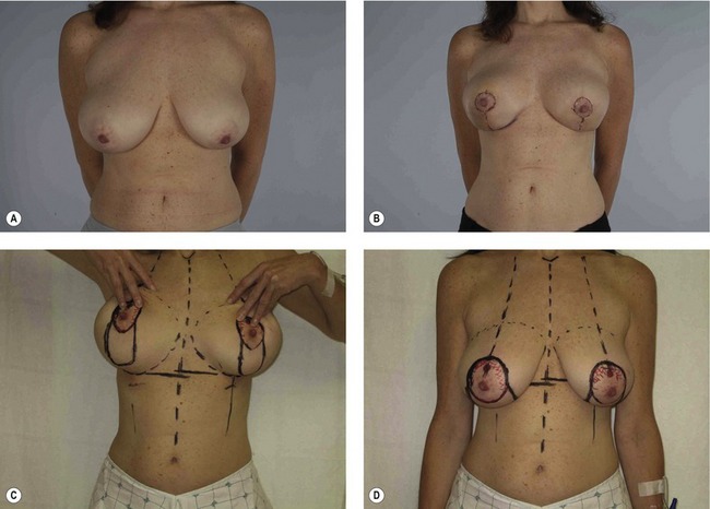

A detailed breast history is required regarding previous breast surgery, current bra size, changes in breast size due to childbirth or breast feeding, family history of breast disease, and mammographic screening (recommended after age 40, but earlier in patients with significant family history). Patient goals need to be clarified. Specifically, volume goals and desired breast shape should be discussed. Patients who desire upper pole fullness and/or volume beyond their current size should consider augmentation in addition to mastopexy. An extensive discussion of risks such as scars, loss of nipple sensation, inability to breast feed, postoperative asymmetry, recurrent ptosis, and nipple necrosis should be included. Implant related risks should also be discussed.


Measurements and assessments as listed above should be performed during the initial consultation. Assessment of the frontal view is critical in terms of decision making. In patients where skin is visible below the areola on frontal view, simple augmentation without mastopexy may be enough to give an excellent result. For patients without skin visible below the areola or for patients that desire more complete elevation of the breast, mastopexy will be required regardless of whether an implant is used. In the massive weight loss patient, ptosis may be so severe that mastopexy with augmentation should be done in two stages.


Marking


The patient is marked while standing, often on the day prior to surgery. Midline, bilateral breast meridians, and inframammary folds are marked (Fig. 48.1). Nipple height is usually set at or just above the level of the inframammary fold in mastopexy patients, but up to 4 cm higher than the fold in mastopexy with augmentation patients in order to center the nipple and breast over the implant. Based around the selected nipple position, a circumareolar pattern is drawn either free hand or using the circular portion of a Wise pattern ring. The superior aspect of the circumareolar marking should be 2 cm above the new nipple position. The lateral, medial, and inferior markings should skirt the areola creating an oval that includes the existing areola.


The breast is then displaced medially and laterally to place the vertical markings in line with the breast meridian (which was marked earlier on the chest wall below the inframammary fold). A horizontal line is then placed to join the vertical markings approximately 2–4 cm above the inframammary fold.


Mar 4, 2016 | Posted by in Aesthetic plastic surgery | Comments Off on Mastopexy with and without augmentation

Full access? Get Clinical Tree

Get Clinical Tree app for offline access