Clinical photography for the aesthetic patient

CHAPTER 2 Clinical photography for the aesthetic patient


The world is filled with forms: objects, people and other things that we look at, identify, enjoy, define, interact with, and make judgments about. When we look at these we are not seeing the things themselves, we are seeing how they interrelate with the light that they are seen in.


The word “photography” means “light writing”. The old saying that “a picture is worth a thousand words” is simply not true; a picture contains more and different information than words could possibly express. The goal of clinical photography is to observe conditions on the body, record them and record changes that happen to them. These recordings may be made for any number of reasons, but for a plastic surgeon the usual aim is to identify pre-existing conditions, modify them and to see how the modifications worked. Pictures are invaluable in the record of patient care for both the surgeon and the patient. They are referred to on each patient visit. They may show the steps to a surgical triumph. At worst they may need to provide a defense in a court of law.


The body exhibits highly complex curvatures and shapes. Though not perceptual psychologists, we are amazed at how selective, limited and evanescent visual memory can be. It is common in our practice to have a patient say that a certain wrinkle “just appeared”, when wrinkles typically take years to emerge. We have noticed this in our own mirror. Many patients, insisting that they know every pore on their faces, for example, will be surprised when a fairly large feature is pointed out to them. With astonishing rapidity, sometimes within days, patients forget how they used to look, after an operation. The surgeon’s visual memory is frequently not much better.


A surgeon’s analytic focus may be on a particular structure while not noticing an adjacent area a centimeter away, and a certain structure or configuration may be present before treatment and not noticed until afterwards. This latter situation is frustrating to patient and surgeon alike as the patient is convinced that there is a “new” problem as a result of surgery. The surgeon cannot prove otherwise without a good visual record. Similarly, areas remote to the surgery may be problematic and without photos there is no good way to analyze them.


The desiderata of clinical photography are straightforward. One must be able to see the conditions present on a print or a screen, and one must see them in the same way at some future time. The images must be consistent over time in lighting and in position. Though simple in concept these are difficult to achieve in practice.


Digital photography has advanced quickly to become the main tool used by plastic surgeons for clinical documentation. Digital imaging is less expensive, more flexible and easier to archive than film, though backup becomes an issue. The resolution of the images continues to improve. Early in the evolution of digital photography, digital point and shoot cameras were popular; however the image quality with small zoom lenses and small image sensors left much to be desired.1


Plastic surgery is a professional endeavor with high standards for record keeping. We prefer the use of digital SLRs (single lens reflex cameras) which use interchangeable lenses with more sophisticated flashes. Though a detailed explanation of CCD sensors, pixel dimensions and print size, and printed dots is beyond the scope of this chapter, we would note that for practical purposes the horizontal resolution (number of pixels of width of a certain image) times the vertical resolution (same) will give the pixel size of the image, usually measured in megapixels. Most commercial printers print at 300 pixels per inch for photographic quality. Thus a camera that has a resolution of 8 megapixels will be able to print at about 8 × 10 inches. Larger prints may be made with some reduction in quality. We think that a 4–5 megapixel range is a reasonable minimum for professional applications.


The use of digital technology allows the taking of large numbers of images with essentially no unit increase in costs. At the time of this writing memory storage costs have plummeted, thus making storage and backup much less expensive than even a few years ago. Patient images may be stored in specialized programs that provide archiving with other functions. An example is the Mirror System from Canfield Scientific.


Lighting


Digital photography is a continuation of traditional photography, and the rules of exposure, lighting and composition are the same as they have been for the last 170 years.


The exploration of light, shadow, form and position has been one of the triumphs of western art. Highly talented people have devoted careers to understanding these relationships and there is a sizeable literature on photography for these ends. Unlike portraiture, where the intent is to capture a face or body in a flattering way, or to reveal an essential truth about someone or something, the goals of clinical photography are not artistic. They should be an unremittingly honest designation of what is there. It is a visual transcription.2


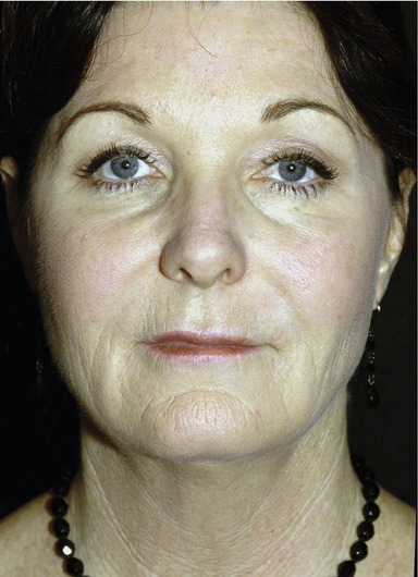
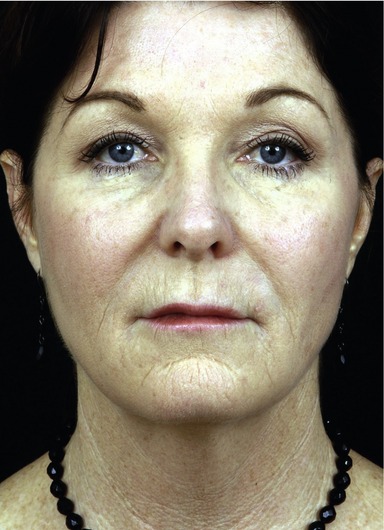
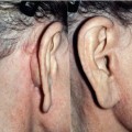
What defines facial and bodily features in the world of daily life is the light one sees them in, or more properly the interplay of light and shadows that visually defines them. Tangential light shows wrinkles, contours and shapes in a very different way than hard anterior light or soft “wrap around” light, which flatten and minimize them (Fig. 2.1).


A recurring problem in the lighting of patient images is that certain details and contours are washed out or flattened so much by the light that they are not visible. Body cellulite or certain facial wrinkles are frequently not seen in anterior (flash) light, but highly visible in everyday vertical light.


In general skin irregularities are best seen in tangential light. Shapes such as breasts, or body contours are best seen in slightly shaded light. Different light shows different things and there is no single light that will show everything, so compromises have to be made. The more attractive a particular lighting setup make the person look, the less likely it is to show the issues of skin and shape that are of interest to patients and surgeons.


Lighting schemes


Many plastic surgery lighting designs have been developed. They vary from on camera flashes of different complexities to external lights in different configurations. Portrait lighting is usually asymmetrical, i.e. the sides of the face or body are lit differently for purposes of interpretation of the subject.2 In general clinical lighting should be symmetrical. If time, space, and temperament of the surgeon allow, we like the use of small silver twin umbrellas, mounted higher than the patient’s eye level. This light is somewhat forgiving, but shows reasonable skin detail and by shifting the lights up or down one can see greater degrees of skin detail. The disadvantage of this lighting is that it casts shadows across the nasolabial fold (NLF) and tends to overexpose the tear troughs (Fig. 2.1).


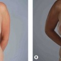
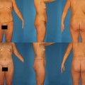
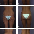
However this lighting scheme is versatile and has enough spread to show body contours well. Even without a dedicated photo room, small slaved flash units with diffusers may be attached to the wall. More vertical light gives very accurate body skin rendition, and so the lights may be elevated or bounced off the ceiling to show skin irregularities for body shots. Light boxes may be used, though we find this light overly flattering. All of these light sources have been used successfully in different offices and all have adherents. The clinical examples shown here use twin umbrella lighting.1


The assumptions made for the purposes of this chapter are that many surgeons do not have the room and inclination to set up external lights and most will use an on-camera flash.

Stay updated, free articles. Join our Telegram channel

Mar 4, 2016 | Posted by in Aesthetic plastic surgery | Comments Off on Clinical photography for the aesthetic patient

Full access? Get Clinical Tree

Get Clinical Tree app for offline access