Abdominoplasty techniques

CHAPTER 66 Abdominoplasty techniques


History


This chapter describes the systematization of a technique developed in the early 1960s and presented in 1964. The original technique was extended and formalized in 1971.


The original version of our abdominoplasty procedure is characterized by low incision, cranial undermining of the flap, musculo-aponeurotic treatment, caudal traction, resection, umbilicus transposition and closure of the wound. However, this type of procedure often results in an asymmetric scar due to irregular traction of the flap. On adipose abdomens, this problem is aggravated by the difficulty in supporting an extremely heavy flap during the resection. In these cases, we shift to performing the block resection of the previously demarcated flap, with lateral dissection, which will facilitate the surgery, provide better control over bleeding, besides reducing intervention time.


As we had felt secure and comfortable in the previous resections of tissue in large abdominoplasties, we began to apply this technique as the preferred method in practically all cases.


The technique, besides facilitating surgery, with all the benefits that have been pointed out, further permits association of abdominoplasty with other surgeries, such as face lifting, mammaplasty–face lifting and thigh lifting. Breast reconstruction using the rectus abdominis muscle, even though it has no link with our technique, represents well the block resection that we have idealized. The results have been consistently satisfactory, corroborated by the surgeons who have used this method.


Physical evaluation


The evaluation presupposes a selection for the diverse variants of the block resection. Technique I is obvious and rarely causes doubts. The other variants require careful reflection, which should be discussed with the patients regarding the benefit, extent of the scars and other consequences. Another factor is whether the abdomen is primary or secondary.


Listening attentively to the complaint and sensing the level of expectation of the patients, the majority female, normally provides a reasonable degree of information regarding the extent of the surgery, its implications and consequences.


It is important to warn about the existence, location and size of the scars during the physical exam. In order to be more objective, it is helpful to draw the supposed scars on the patient’s abdomen.
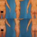

The patient should be examined in a standing position with abdomen exposed. The seated exam should be added when such would provide greater clarity. In this position, the patient has difficulty in contracting the abdominal musculature, which facilitates real knowledge of the shape of the abdomen, showing the situation in its fullest degree.


After the exam, all the implications of an abdominoplasty are discussed exhaustively: age, fertility, time necessary for recovery, return to his/her normal activities, period of internment, type of anesthetics, dressings, drains, as well as risks arising from the surgery.


The most frequent complication must be pointed out, namely deep vein thrombosis in the lower limbs, and factors that predispose the patient to this incident should be discussed. It is also opportune, although it may seem premature, to instruct the patient about what measures are necessary for prophylaxis of the DVT.


It is important to associate an abdominal ultrasonography to the basic exams, so as to detect any pathology that could contraindicate the surgery.


Women of child-bearing age should be warned that abdominal stretch with future pregnancies would interfere with the quality of the result.


In secondary abdomens with the presence of marked irregularities and supposition of hernia, it is also appropriate to use magnetic resonance.


In abdomens with large distension or voluminous incisional hernias, the wearing of a girdle for abdominal containment for around a month prior to surgery is fundamental. Thus, a gradual adaptation will occur after surgical adjustment of the abdominal wall, as the consequent elevation of the diaphragm could entail respiratory dysfunction in the postoperative period.


It is of great importance to know all the medication normally used, emphasizing the need to suspend those substances that could interfere with coagulation two or three weeks before surgery.


Smokers represent a serious potential for the flap to incur damage. Even those who affirm that they have given up smoking in time (minimum one month prior to surgery) are considered smokers, requiring the care inherent to this type of situation.


Anatomy of the abdominal wall
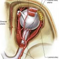

The abdomen is the focus of multiple complaints by patients seeking plastic surgery. Due to its evident location, modifications to the form and volume produce, at times, unpleasant alterations to the corporal aesthetics. The abdominal protrusion can be associated to diverse factors such as adiposity, flaccidity of skin and musculature, hernias, increase in intra-abdominal volume, or a combination of these elements.


The anterior abdominal wall presents lines that serve as anatomical references. The medial anterior midline of the abdomen, the linea alba, is a point where the subcutaneous layer is diminished, and is often practically absent in lean patients, with defined musculature. The alba line serves as a reference for the correct positioning of the incisions during surgery of the abdomen. It is formed by the junction of the aponeuroses of the rectus abdominis muscle in the medial line, and extends from the xiphoid process to the pubic symphysis; is broader in the upper portion and suffers alterations with the increase in intra-abdominal volume, as in pregnancy, ascites and obesity. The linea semilunares indicate the lateral edges of the rectus abdominis muscle, and the tendinous inscriptions form transverse folds, three on average.


The disposition of fat varies according to gender. We know that, in females, fat tends to form pockets and maintains a predominant disposition in the infra-umbilical portion of the abdomen, whereas, in men, the distribution is more generalized.


Vessels of the abdominal wall


The blood supply is formed of two primary arterial plexuses: a superficial subdermic system and a deep aponeurotic muscle.


Small arteries originate from the anterior and collateral branches of the posterior intercostal arteries of the intercostal spaces 10 and 11 and of the anterior branches of the subcostal arteries to supply the muscles in the antero-lateral region. They are anastomosed with the superior epigastric arteries, with the superior lumbars and among themselves.


The main arteries of the antero-lateral abdominal wall are the lower epigastric and deep circumflex iliac, branches of the external iliac artery, associated to the superior epigastric artery, which is the terminal branch of the internal thoracic artery.


The superficial epigastric and lateral thoracic veins are anastomosed, thereby joining the veins of the upper and lower halves of the body. The three superficial inguinal veins terminate in the magna saphena vein of the lower limb. The upper and lower epigastric vessels run together from the posterior face of the rectus abdominis muscle, inside its sheath.


Lymphatic drainage is carried out by two networks, one superficial and the other deep. The superficial lymphatic vessels from the upper abdominal wall to the umbilicus continue on to the axillary lymph nodes, and the lower drain to the inguinal lymph nodes. The lymph of the musculature is also drained to the deep lumbar and iliac lymph nodes.


The lymphatic drainage necessarily runs to the axillary nodes in the immediate postoperative period of abdominoplasty due to the section of lymphatic canals that drain the inguinal nodes. The systems tend to rebuild themselves as time goes by.


Mar 4, 2016 | Posted by in Aesthetic plastic surgery | Comments Off on Abdominoplasty techniques

Full access? Get Clinical Tree

Get Clinical Tree app for offline access