In many cases of complex facial defects, because of advanced cutaneous malignancies, primary wound closure is impossible. In these instances, ideal results can be obtained through recruitment of adjacent tissue with the use of local flaps. Advances in local flap techniques have raised the bar in facial reconstruction; however, acceptable results to the surgeon and patient require high levels of planning and surgical technique. Defects resulting from Mohs surgery and other traumatic injuries can typically be repaired with local flaps. A well-planned and executed local flap can lead to excellent cosmetic results with minimal distortion of the surrounding facial landmarks.
Key points
- •
Local facial flaps offer a good option for repair of Mohs micrographic surgery for cutaneous lesions.
- •
Rotation flaps are curvilinear in nature and rotate adjacent tissue into a defect.
- •
Transposition flaps are linear and pivot toward a defect over an incomplete bridge of tissue.
- •
Rhombic and bilobe flaps incorporate components of transposition and rotation flaps and serve as the workhorse flaps of Mohs reconstruction.
- •
Local facial flaps are hearty, only suffering rare, mild complications.
Background
Modern evolution in techniques of facial reconstruction have dramatically increased the possibilities for repair of facial defects. The need for advanced facial reconstruction has grown significantly since the advances of Mohs micrographic surgery, which represents the gold standard for malignancies of the face and neck. Use of immediate fresh tissue fixation allows for Mohs surgical excisions to be performed quickly, facilitating expedient repair. In many cases of complex facial defects resulting from the extirpation of advanced cutaneous malignancies, primary wound closure is impossible. In these instances, ideal results can be obtained through recruitment of adjacent tissue with the use of local and regional flaps. Advances in local flap techniques have raised the bar in facial reconstruction; however, acceptable results to both the surgeon and the patient require high levels of planning and surgical technique.
Defects resulting from Mohs surgery and other traumatic injuries can typically be repaired with grafts or local flaps. Between these options, local flaps are often preferred because of their superior color match and texture. A well-planned and executed local flap can lead to excellent cosmetic results with minimal distortion of the surrounding facial landmarks. Local flaps used for facial reconstruction are classified by a variety of methods, including blood supply, flap contents, and the method of transfer. Rotation flaps are curvilinear flaps that pivot into the defect. Transposition flaps are linear and pivot toward the defect over an incomplete bridge of skin. This is in contrast to interpolation flaps, which pivot toward defects over intact bridges of skin. The rhombic flap, bilobe flap, O-T/O-Z flap, and note flap are types of transposition flaps, some of which include both transposition and rotation components.
Rotation flaps
Rotation flaps are designed with curvilinear orientation in the direction of the defect that they pivot toward. Although these flaps are rotational in their direction, they also span the defect by stretching the elastic tissues. This leads to the points of greatest wound closure tension occurring along the distal border of the flap rather than along the length of the flap. The secondary defect that occurs following execution of a rotational flap is determined by the size of the flap, with a larger-rotation flap leading to a narrower and longer secondary defect. A narrower secondary defect will lead to less tension on closure of the wound; however, this exists only up to a certain point. Larrabee and Galt demonstrated there is minimal benefit to extending the arc of rotation flaps beyond 90° from the axis of the defect.
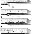
The ideal defect for rotation flaps are triangular in shape. The height-width ratio of the triangle ideally should be 2:1. The arc of rotation extending from the base of the triangle should be a symmetric curve, with the radius of the curve being 1 to 2 times the height of the triangle ( Fig. 1 A). For optimal results with rotation flaps, the defects can be modified into triangular shapes via conservative excision of normal tissue. Burow triangles are often used to assist with closing the secondary defect. The length of the flap should be 4 times the width of the base of the triangular defect (see Fig. 1 B, C). This ratio obviates the need for excision of a Burow triangle to equalize the defects. Enlarging the flaps beyond the 4:1 ratio does not significantly decrease the wound closure tension; however, a longer flap can be useful in areas of limited skin mobility.
Wide undermining is performed to allow for pivoting of the flap toward the defect. Undermining can also reduce the extent of standing cutaneous deformity by shifting the deformity slightly away from the flap’s base. When a standing cutaneous deformity persists in spite of these techniques, secondary excision of the deformity is warranted. In most instances, a standing cutaneous deformity will flatten over 6 weeks postoperatively. Additionally, close attention must be paid to the closure of deep layers, ensuring close approximation at the points of maximal tension and meticulous eversion of the skin edges.
Rotation flaps have several significant advantages in the repair of facial defects. There is a great amount of flexibility in the planning and orientation of the arc of the flap. This can allow for orientations that promote lymphatic drainage, and also that minimize vascular and nervous disruption. The flap is very robust with strong vascular flow due to its broad-based design. The surgeon can also often position the single long arc of the flap within a relaxed skin tension line (RSTL) or aesthetic unit for greater scar camouflage.
The rotation flap has utility for many types of facial defects; however, because of the curved incision of the flap, it sometimes does not lie cleanly within an RSTL. In cases in which the incision does not lie within an RSTL, the scar can be less camouflaged and more noticeable than in other methods of repair. Because of the degree of rotation, these flaps often develop standing cutaneous deformities at their base. These deformities cannot be initially excised, as that would compromise the vascular supply to the tissues.
Rotation flaps are not the optimal choice in repair of central cheek defects or most nasal defects. In men, the rotation flap can distort the hair-bearing skin of the sideburn medially toward the malar eminence. The skin of the nasal tip is very inelastic, making recruitment difficult. Additionally, the donor site scars along the nose do not typically fall cleanly between the nasal aesthetic subunits. Most nasal defects are better repaired with other flaps, such as the bilobe or rhombic flap.
Bilobe flap
The bilobe flap is generally considered one of the “workhorse” flaps of facial reconstruction. Classified as both rotational and transpositional, the bilobe flap is a transposition flap because it is elevated and mobilized toward an adjacent defect and transposed over an incomplete bridge of skin, and a rotational flap because it pivots around a specific point and maintains its radius. It is particularly useful when a single transposition flap exerts too much tension on the closure. The bilobe flap is able to more effectively transfer tension across a greater angle of rotation and therefore distributes the load both more equally and with a larger component being placed away from the primary defect.
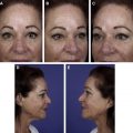
Originally described for nasal tip reconstruction by Esser in 1918, the bilobe flap continues to serve the facial plastic surgeon during post-Mohs micrographic surgery defect repair. Although the use of the bilobe flap can be used in many parts of the body, its utility is greatest in areas that need to minimize tension in the area directly adjacent to the primary defect, like the nasal dorsum, sidewall, and tip subunits ( Fig. 2 ).
As described previously, this flap is best used for defects of the nasal tip, dorsum, and sidewall equal to or less than 1.5 to 2.0 cm. Extension to the nasal ala reduces this flap’s efficacy as the scarring that occurs will many times cause cephalic elevation, retraction, and/or distortion of the alar rim leading to lesser cosmetic outcomes. The Zitelli-modified bilobe flap is designed with a total arc of rotation 100° or less, with each limb of the flap rotating 45 to 50°. The flap is routinely based medially along the nasal dorsum or laterally along the nasofacial groove, with laterally based flaps resulting in better concealed scars. The bilobe flap is most successful when the skin is elevated in the subcutaneous plane and the raised lobes closely match the thickness of the defect area. When there is a considerable mismatch, a delayed closure may be beneficial by allowing a deeper defect time to granulate in its base. This allows for a flap that may not need much alteration and produces a more even level to the healed flap and surrounding skin.
When designing a bilobe flap, first measure the radius of the defect. Once obtained, mark a point to the side of the defect, preferably laterally, equal to the radius of the defect. Next, measure 2 arcs: one equal to 2 times radius and the other 3 times the radius, spanning the entire area of the defect and planned area for flap rotation. Once the 2 arcs are marked, use the diameter distance of the defect to measure the base of the first lobe along the first arc immediately adjacent to the defect, ensuring the middle of the lobe is 45 to 50° from the center of the defect. The height of the first lobe should extend to the line of the second arc, therefore making its height equal to the radius of the defect. Next, again measuring along the first arc line, the second lobe should be a distance slightly smaller than the first lobe, and again 45 to 50° from the middle of the first lobe or 90 to 100° away from the middle of the defect. Its height is a distance twice that of the first lobe, and of a slightly more triangular shape. Finally, mark the standing cutaneous deformity between the edge of the defect and the initial mark placed equal to 1 radius of the defect ( Fig. 3 ).